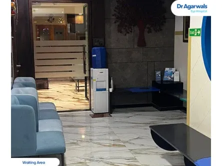
Dr Agarwals Eye Hospital - Thane, Maharashtra
Address
1st floor, 102, Soham Plaza, Tikuji Ni Wadi Rd, near Manpada Flyover, next to Titan Hospital, Manpada Naka, Manpada, Thane West, Thane - 400607
Opening Hours
Mail ID
care@dragarwal.com
Phone
Eye Treatments Available at Dr Agarwals Eye Hospital
Nuclear Cataract
What is Nuclear Cataract?
An excessive amount of yellowing and light scattering affecting the centre of the lens is called nuclear cataract. The nuclear sclerosis is when the nucleus, i.e. the center of the eye, begins to get cloudy, yellow, and hardens. A part of the aging process in humans, nuclear sclerotic cataract also occurs in dogs, cats, and horses. When nuclear sclerosis eyes worsens, i.e. the lens becomes cloudier with age, the condition is called nuclear cataract. Further dehydration of the nucleus and the cortical part of the lens, coupled with heightened sclerosis, leads to nuclear senile cataract.
Sometimes, a cloudy lens might be present at birth, which is called a congenital cataract. When the congenital cataract is present near the nucleus of the eye, it is called congenital nuclear cataract or fetal nuclear cataract.
Nuclear cataract symptoms
Nuclear cataract affects distance vision. Thus, anything that involves seeing things at a distance would prove difficult. Other symptoms for nuclear cataract could include:
- Difficulty in driving, reading signboards
- Occasional double vision
- Difficulty reading things at a distance
- Severe glare from lights
Nuclear cataract risk factors
While age is the predominant factor for the development of nuclear cataract, the following can also be considered as risk factors for nuclear cataract
- Smoking
- Increased exposure to UV light
- Steroid use
- Diabetes
How to diagnose a nuclear cataract?
A number of tests can help the doctor diagnose a patient with a nuclear cataract. The tests are:
- Dilation: The doctor administers drops into the eye of the patient, which will dilate the retina of the eye. This opens up the eye and helps the doctor examine the interior of the eye, including the lens.
- Slit lamp test: The doctor uses a special microscope-like device with a light on it to examine different parts of the eye – the cornea, iris, and the lens, including the nucleus of the lens.
- Red reflex test: The doctor bounces the light off a surface and uses a special magnifying glass to examine the eye in the reflection of this light. When the eyes are healthy, they appear red in this test.