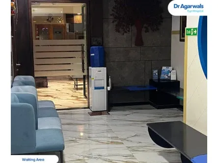
Dr Agarwals Eye Hospital - Thane, Maharashtra
Address
1st floor, 102, Soham Plaza, Tikuji Ni Wadi Rd, near Manpada Flyover, next to Titan Hospital, Manpada Naka, Manpada, Thane West, Thane - 400607
Opening Hours
Mail ID
care@dragarwal.com
Phone
Eye Treatments Available at Dr Agarwals Eye Hospital
Angle Recession Glaucoma
Angle Recession Glaucoma: Meaning, Symptoms & Treatment Explained
Angle recession glaucoma is a type of secondary glaucoma that develops after eye trauma, particularly blunt injuries. It occurs when the internal drainage structures of the eye are damaged, leading to increased intraocular pressure (IOP) and gradual optic nerve damage.
This condition can appear months or even years after the original injury, making long-term monitoring crucial. Understanding its meaning, causes, and treatment options helps in early detection and prevention of permanent vision loss.
What is Angle Recession Glaucoma?
Angle Recession Glaucoma Meaning
Angle recession glaucoma is a condition caused by structural damage to the anterior chamber angle of the eye. In this region, fluid drains through the trabecular meshwork.
When blunt trauma occurs, this angle separates or “recedes,” leading to reduced drainage efficiency and higher intraocular pressure. Over time, this persistent pressure damages the optic nerve, resulting in vision loss. Although not every trauma leads to glaucoma, people with significant angle damage remain at lifelong risk.
How Angle Recession Glaucoma Develops
Angle recession glaucoma develops after blunt trauma, causing tears or separation within the ciliary body and trabecular meshwork. This structural injury compromises the eye’s outflow of aqueous humour, allowing aqueous humour to accumulate and raise IOP.
The process may remain silent for months or years before symptoms emerge. In some cases, the eye initially heals from the trauma but later develops angle recession glaucoma due to gradual trabecular degeneration and scarring, which impede normal drainage.
Causes and Risk Factors of Angle Recession Glaucoma
Eye Trauma and Blunt Injuries
The leading cause of angle recession glaucoma is blunt eye trauma. Injuries from sports activities, motor vehicle accidents, or physical assaults can generate enough force to damage the drainage angle. Objects like balls or fists striking the eye are particularly common causes.
Additional Risk Factors
Certain factors increase susceptibility, including older age, previous ocular injuries, and pre-existing conditions such as uveitis or high myopia. The extent of the initial trauma often determines the severity of angle recession glaucoma, with deep-angle tears posing a higher risk.
Symptoms of Angle Recession Glaucoma
Early Symptoms to Watch For
The angle recession glaucoma symptoms can be subtle in the early stages. Patients may experience occasional blurred vision, mild eye discomfort, or halos around lights.
Because pressure build-up progresses slowly, many remain asymptomatic until significant optic nerve damage occurs. Regular follow-up after eye trauma is essential to detect early signs.
Advanced Symptoms
In later stages, peripheral vision loss becomes noticeable, often described as “tunnel vision.” Patients may find it difficult to see objects at the edges of their visual field. Without timely angle recession glaucoma treatment, vision may progressively deteriorate, leading to permanent impairment.
Diagnosis of Angle Recession Glaucoma
Eye Examination Techniques
Diagnosis begins with a comprehensive ophthalmic evaluation. Tonometry measures intraocular pressure, while gonioscopy allows direct inspection of the anterior chamber angle to confirm recession or scarring.
A slit-lamp examination helps assess any corneal, iris, or lens changes following trauma. Together, these tests establish the extent of structural damage and its impact on eye pressure.
Imaging and Additional Tests
Advanced imaging techniques such as optical coherence tomography (OCT) and visual field testing are used to detect early optic nerve changes and monitor disease progression. These tools provide detailed images of the drainage structures and nerve fibre layers, allowing precise assessment of angle recession glaucoma severity.
Post-Treatment Care and Monitoring
Post-treatment care focuses on consistent follow-ups to evaluate IOP, optic nerve health, and visual fields. Patients should use prescribed eye drops as directed and avoid activities that risk further trauma. Wearing protective eyewear during sports or hazardous work is essential to prevent recurrent injury and worsening of angle recession glaucoma.
Prognosis and Long-Term Outlook
Angle recession glaucoma is a chronic condition requiring lifelong monitoring. The prognosis depends on early detection and adherence to treatment.
With proper management, most patients can maintain stable vision. However, untreated cases can lead to irreversible optic nerve damage. Regular eye examinations after trauma are vital for preventing severe visual loss.
Conclusion
Angle recession glaucoma is a secondary form of glaucoma caused by blunt eye trauma that damages the eye’s drainage angle. It can develop long after the initial injury, making periodic eye evaluations essential for early diagnosis.
Recognising angle recession glaucoma symptoms and accessing timely treatment can preserve sight and prevent long-term complications. Anyone who has sustained an eye injury should consult an ophthalmologist for a detailed assessment and continued monitoring.