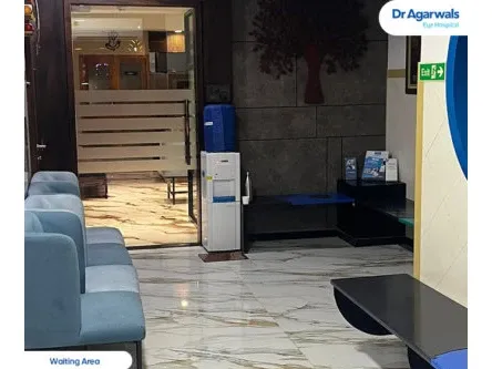
Dr Agarwals Eye Hospital - Thane, Maharashtra
Address
1st floor, 102, Soham Plaza, Tikuji Ni Wadi Rd, near Manpada Flyover, next to Titan Hospital, Manpada Naka, Manpada, Thane West, Thane - 400607
Opening Hours
Mail ID
care@dragarwal.com
Phone
Eye Treatments Available at Dr Agarwals Eye Hospital
Pterygium or Surfer’s Eye
Pterygium or Surfers Eye Treatment
What Is a Pterygium
A pterygium is a fleshy, triangular growth of tissue that extends across the white of the eye toward the cornea, often linked to prolonged sun and wind exposure.
When It Becomes a Concern
While often harmless initially, growth toward the cornea can cause irritation, redness, and distorted vision if left untreated.
Effective Surgical Removal
Our surgeons remove the growth using techniques designed to minimise recurrence, helping restore comfort and a clear, natural eye surface.
Protective Habits Going Forward
Alongside treatment, our team offers guidance on sun protection and eye care habits that can help reduce the likelihood of a pterygium returning after removal.
Our Diagnostic Approach
A careful eye examination helps distinguish a pterygium from other surface growths, ensuring the right treatment path is chosen from the outset.
What is Pterygium (Surfer’s Eye)?
Pterygium, commonly known as Surfer’s Eye, is a non-cancerous growth of tissue on the white part of the eye (conjunctiva) that can extend onto the cornea. It often develops in individuals frequently exposed to UV rays, wind, and dust—hence the name “Surfer’s Eye.” This condition can cause discomfort, vision disturbances, and, in severe cases, interfere with eyesight.
Although pterygium is not life-threatening, it can be progressive, requiring medical intervention in some cases. Early detection and management can prevent complications, making awareness essential for individuals at risk.
Symptoms of Pterygium (Surfer’s Eye)
Pterygium symptoms vary depending on its size, growth rate, and location. Common signs include:
- Foreign Body Sensation
A persistent feeling of something stuck in the eye is common, leading to discomfort, excessive blinking, or rubbing.
- Tearing from the Eyes
Excessive tearing, also called epiphora, occurs as the eye tries to remove irritation caused by the growth.
- Dryness of the Eyes
Pterygium disrupts the normal tear film, leading to chronic dry eyes, discomfort, and irritation.
- Redness
Due to inflammation, the affected eye often appears red and irritated, especially after exposure to wind or sunlight.
- Blurred Vision
As the pterygium extends onto the cornea, it can distort vision, leading to blurred or obstructed eyesight.
- Eye Irritation
A growing pterygium can cause itching, burning, and overall discomfort, especially in dry or dusty environments.
Causes of Pterygium (Surfer’s Eye)
Pterygium is strongly linked to environmental factors, especially exposure to UV rays, wind, and dust. Other contributing factors include:
- UV Exposure (Biggest Risk Factor)
Prolonged exposure to ultraviolet (UV) radiation from the sun is the primary cause of pterygium. This explains why it is commonly seen in surfers, fishermen, and outdoor workers.
- Chronic Eye Irritation
Frequent exposure to dry, dusty, or smoky environments increases irritation, making individuals more susceptible to pterygium.
- Wind and Sand Exposure
High-wind environments, especially in coastal areas, can lead to increased evaporation of tears, making the eyes dry and prone to irritation.
- Genetic Factors
Studies suggest that genetics may play a role in pterygium development, with some individuals being more prone to the condition.
- Contact Lens Use & Eye Strain
Excessive contact lens wear or digital eye strain may contribute to discomfort and increase the risk of pterygium self-care negligence.
Types of Pterygium (Surfer’s Eye)
There are different grades of pterygium based on severity:
Grade 1: Small growth without affecting vision
Grade 2: Larger growth but no major vision obstruction
Grade 3: Pterygium extends over the cornea, causing mild vision impairment
Grade 4: Severe obstruction, leading to blurred vision and astigmatism
Best Treatment Options for Pterygium (Surfer’s Eye)
1. Conservative Management (Self-Care)
For mild cases, treatment involves:
- Lubricating eye drops for dryness relief
- Anti-inflammatory drops to reduce redness and irritation
- UV-blocking sunglasses to prevent further damage
2. The Best Eye Drop for Pterygium
Some of the best-recommended eye drops for pterygium include:
- Artificial tears (for dryness relief)
- Steroid eye drops (for inflammation)
- Lubricating gel-based drops (for overnight hydration)
3. Surgical Removal (For Advanced Cases)
In cases where the pterygium affects vision, surgical removal is recommended. The procedure involves excising the growth and using a conjunctival autograft to prevent recurrence.
Prevention Tips: How to Reduce the Risk of Pterygium?
- Wear UV-Protective Sunglasses – Reduces UV exposure
- Use Lubricating Eye Drops – Prevents dryness and irritation
- Limit Outdoor Exposure – Avoid peak sun hours (10 a.m. – 4 p.m.)
- Wear Hats & Protective Eyewear – Acts as an extra barrier
- Stay Hydrated – Keeps eyes moist and reduces irritation
By following these pterygium self-care tips, you can minimize the risk of developing this eye condition.
Tests for Diagnosing Pterygium (Surfer’s Eye)
Early detection of pterygium (Surfer’s Eye) is crucial in managing the condition effectively. Eye specialists use several diagnostic tests to assess the growth, impact, and severity of the condition. Here are the primary tests used for diagnosing pterygium:
1. Slit Lamp Examination
A slit lamp examination is one of the most common tests used to detect pterygium. It allows ophthalmologists to examine the eye’s surface, cornea, and conjunctiva under high magnification and illumination. This test helps assess inflammation, vascular growth, and potential corneal involvement.
2. Visual Acuity Test
A visual acuity test determines how well a person can read letters on an eye chart. As pterygium progresses, it may affect vision clarity by altering the shape of the cornea, causing astigmatism or leading to a foreign body sensation in the eye.
3. Corneal Topography
Corneal topography is an advanced imaging technique that maps the curvature of the cornea. Since pterygium can cause corneal distortion, this test helps identify irregularities and assess how the growth impacts light refraction and vision quality.
4. Photo Documentation
To track the progression of pterygium over time, doctors use photo documentation. This involves taking detailed images of the eye at different intervals to measure growth, monitor changes, and determine if surgical intervention is necessary.
Complications of Pterygium (Surfer’s Eye)
Although pterygium is typically a benign eye condition, complications can arise if the growth progresses unchecked. Here are some potential complications:
1. Chances of Infection
Pterygium increases the risk of eye infections, especially if the surface of the conjunctiva becomes irritated or inflamed. If left untreated, infections can lead to pain, discharge, and further damage to the cornea.
2. Corneal Scarring
A growing pterygium can extend into the cornea, leading to scarring. This scarring can result in permanent vision impairment, as it distorts the natural shape of the cornea and causes blurry vision.
3. Retinal Detachment (Rarely)
Though rare, severe cases of pterygium can contribute to retinal detachment due to chronic inflammation and ocular surface instability. This is a serious condition that can lead to vision loss if not treated promptly.
4. Conjunctival Graft Dehiscence
In cases where a pterygium surgery (conjunctival autograft) is performed, there is a possibility of graft dehiscence, meaning the transplanted tissue fails to adhere properly, requiring further intervention.
5. Diplopia (Double Vision)
When pterygium grows aggressively, it can cause diplopia (double vision) due to its effect on corneal curvature and visual alignment. This can severely impact daily activities such as reading, driving, and working on screens.
Treatment for Pterygium Eye
1. Medical Treatment
Mild cases of pterygium can be managed with non-surgical treatments, including:
- Lubricating eye drops (Artificial tears) to reduce dryness and irritation
- Anti-inflammatory eye drops to control redness and swelling
- UV protection (sunglasses and hats) to slow progression
- Lifestyle modifications, such as reducing dust and sun exposure, can also help prevent worsening symptoms.
2. Surgical Treatment
For advanced cases where pterygium obstructs vision or causes severe discomfort, surgical removal is required. The surgical procedure involves:
- Excision of the pterygium to remove the growth
- Conjunctival autograft transplantation, where tissue from another part of the eye is placed over the affected area to prevent recurrence
- Amniotic membrane transplantation in severe cases to promote healing and reduce scarring
Post-surgery, regular follow-ups and UV protection are essential to prevent recurrence.