
Cylindrical Sight : Causes, Symptoms, and Correction Methods
May/28/2026
Address
E-2/66 Arerra Colony, 10 No. market,
Arera Colony,
Bhopal - 462016
Mail ID
care@dragarwal.com
Behcet’s Disease
Behcet’s Disease, also called Silk Road Disease, is an autoimmune disease in which the blood vessels of your body get inflamed (a defense reaction of your body to any stimulus).
Below we have mentioned some of the many symptoms of Behcet’s disease:
A group of four symptoms are commonly known to occur in this disease: Mouth ulcers, Genital Ulcers, Skin problems and Inflammation inside your eye. Your joints, digestive system and nervous system may also be affected.
Inflammation inside your eyes can cause uveitis (uvea is the area around your pupil), retinitis (retina is the light-sensitive tissue in your eye) and iritis (iris is the coloured part of your eye).
What causes your own body’s cells to attack the blood vessels is not exactly known. People of Asian and Eastern Mediterranean origin are found to suffer more frequently. Men are affected more commonly than women and especially so during their 20s and 30s. Genetic factors combined with environmental factors like microbes are thought to play a role.
There is no specific cure for this disease. However, when it comes to treatment for behcet’s disease, it consists of medications to reduce your discomfort, control the Inflammation of your and prevent severe complications. Medications include steroids to suppress the errant immune system, colchicine etc. Steroid eye drops and steroid injections next to your eye may be given.
This Behcet syndrome triad is marked by its long term duration and recurrence. However, you may have periods when you go into remission (your symptoms go away temporarily). The severity of your disease may vary from you even leading a normal life to becoming blind and severely disabled. Vision loss may be kept in control by keeping the disease in remission.
Cataract
An eye cataract forms when the lens of the eye becomes cloudy, making it harder to see clearly. The lens, which is normally clear, helps focus light on the retina to produce sharp vision. When it becomes cloudy, it can cause cataract symptoms like blurry vision, glare, and trouble with night driving. Although common in older adults, eye cataracts can also be caused by injuries, medical conditions, or prolonged UV exposure. Cataracts progress slowly but can be effectively treated with modern surgical techniques.
Cataract symptoms vary depending on the type and stage of the cataract. Common cataract signs and symptoms in the eye include:
There are several causes of cataract, with aging being the most common. Other factors that lead to the formation of cataracts include:
The risk of developing cataracts increases due to several factors. Common cataract risk factors include:
While not all cases of cataracts can be prevented, adopting healthy habits can help delay their onset. Here’s how you can reduce your risk of developing eye cataract symptoms:
To ensure proper healing after cataract surgery, follow these tips:
Most patients experience improved vision within a week, with full recovery in 4-6 weeks.
For a safe and effective medical treatment of cataract, Dr Agarwals Eye Hospital is a one-stop solution. We provide a safe eye cataract treatment based on the type of cataract, including cortical cataract, intumescent cataract, nuclear cataract, posterior subcapsular cataract, rosette cataract, and traumatic cataract. We also provide paediatric cataract treatment and effectively cater to complicated cataract treatment.
Reach out to our team for thorough analysis, treatment options, and prevention tips!
The eye care specialists of our hospital diagnose cataract with a comprehensive eye examination. To identify cataract, your eye doctor analyses your medical history. If you experience vision difficulties, they also look for such signs and symptoms through some tests before cataract treatment that include:
For better examination of your eyes, eye specialists use eye drops to widen your pupil. It allows them to get a closer view of your retina.
With an ophthalmoscope, eye doctors look for the visible signs of cataract and proceed with the treatment accordingly.
In this eye examination, your eye doctor uses an eye chart to understand your vision and ability to read letters from a distance. They perform this test individually on each eye with one eye covered and similarly on the other. If they diagnose any signs of cataract, they proceed with the suitable cataract treatment.
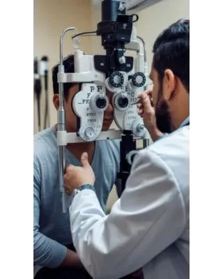
A slit lamp is an instrument with a high-intensity light beam that allows them to see the structures of your eyes better under magnified glasses. They examine the cornea, lens, iris, and other parts of your eyes. With this slit lamp, eye doctors even analyse the small sections, making it easier to detect minor problems.
Cataract is a common eye problem and people get this problem as they age. When you start realising its symptoms, contact the experts of Dr Agarwals Eye Hospital for early cataract treatment. Here are cataract treatment options:
In the initial stage, when you don’t have any vision difficulty, your eye doctor prescribes eyeglasses to correct your vision.
If the symptoms of cataract start impacting your day-to-day chores, then cataract eye surgery is the only effective option for cataract treatment to root out its symptoms. This surgery is also effective for congenital cataract treatment.
When eye doctors determine that your cataract is dense and find difficult to create an opening, they rely on laser treatment for cataract.
When you visit Dr Agarwal’s Eye Hospital, our doctors discuss with you and acquaint you with the cataract operation procedure of working.
In the traditional cataract treatment procedure, eye care specialists numb the area around your eyes with local anaesthesia before the cataract eye surgery, but you are awake throughout. Under this cataract operation, eye surgeons remove the clouded lens using a microsurgical instrument and replace it with an artificial intraocular lens (IOL).
There are here some laser-assisted surgery options to restore your vision:
For the cataract treatment, doctors make an incision through femto laser cataract surgery to access and remove the cataract from your eyes.
The surgeon creates a precise surgical plane for the corneal incision. It is done with a sophisticated 3-D image eye image called an OCT scan. Doctors aim to create an incision in a specific location with accurate depth and length in all planes. With the OCT image and a femtosecond laser, it can be performed exactly.
The blurriness of vision occurs after years later because the eye’s lens capsule gets cloudy. This capsule holds the IOL in its original position. To open this cloudy capsule, doctors may use a laser, which helps you restore your vision. This process of cataract treatment is called capsulotomy.
Under laser cataract surgery, your provider uses a laser for better precision to remove the affected lens for IOL cataract surgery. Once they create an opening, this laser beam triggers the cataract to soften and fragment it easily. This process of cataract treatment is done with help of phacoemulsification probe using ultrasound & mechanical energy.
If your cataract gets hard, it may require more energy. This may increase the chances of more collateral tissue damage as compared to soft cataract. However, our surgeon takes all necessary precautions to lower such tissue damage and perform cataract eye surgery carefully.
During the cataract operation, patients feel little to no pain. To recover faster after a cataract operation, here are some tips:
Since cataract is an age-related problem, you can follow the below cataract precaution tips to protect your vision:
We at Dr Agarwal’s Eye Hospital provide comprehensive treatment for various eye diseases. The diseases are listed here:
To treat your eye-related problems, our eye treatment or surgery options include the following:
Photorefractive Keratectomy (PRK)
Implantable Collamer Lens (ICL)
Retinal Laser Photocoagulation
If you experience blurry vision or glares around lights, immediately schedule your appointment with the eye doctors of Dr Agarwal’s Eye Hospital! With an in-depth examination of your eyes using state-of-the-art facilities, we are committed to offering the best eye care treatment.
Note: Eye cataract operation costs may vary depending on the treatment you seek for. Book your consultation with us today for the best cataract treatment!
Conjunctivitis
Inflammation of the conjunctiva (Transparent membrane covering white part of the eye) is called Conjunctivitis. It is a condition where the eye turns red. Allergic conjunctivitis is caused due to allergy. The agents which cause allergies are known as allergens. Every individual is allergic to one or other substance in the environment. The most common allergens are dried grass, pollen grains etc. List of allergens are endless and individual specific. When an individual who is prone for allergy; get exposed to allergens, it causes release of some chemicals in tissue e.g. Histamines by allergy mediating cells like Mast cells. It causes itching, redness, and watering from eyes. Allergic conjunctivitis is not contagious unlike the traditional red eye or infective conjunctivitis.
Below we have mentioned some of the many signs of allergic conjunctivitis:
Routine examination by an eye doctor is sufficient. Some signs are very specific for allergic conjunctivitis like papillae, ropy discharge, limbal hyperplasia. To find out specific allergens, Allergy test can be done in individuals who are prone for generalized systemic allergy like asthma, eczema, Atopy etc. Otherwise, such tests are not recommended as avoidance to these allergens is practically cumbersome in routine life.
Before getting to allergic conjunctivitis treatment, it is imperative to understand that complete cure of allergy is not feasible, but symptoms of allergy can be suppressed with the help of medicines. Rubbing of eyes due to itching causes more trouble to eyes than allergy itself, so intense rubbing of eyes should be avoided.
Avoidance of allergens is the IDEAL treatment but it’s easier to say than done as it will severely hamper lifestyle and quality of life. How long the allergic conjunctivitis lasts depend on the type, severity and the treatment taken along with the compliance for the treatment.
Medicines in the form of eye drops like mast cell stabilizers (Olopatadine, Sodium Cromoglycate), antihistamines (Ketotifen, Bepotastine), NSAID ( ketorolac), Steroids (loteprednol, FML, Difluprednate, Prednisolone etc), immune modulators (Cyclosporine, Tacrolimus eye ointment), are useful in treating allergic conjunctivitis.
Any eye drops should not be started without an ophthalmologist's opinion to avoid its side effects.
Using sunglasses while going out, cold compression can alleviate allergic symptoms and serve as a useful home remedy for eye itching.
Cornea Transplantation
A corneal transplant involves surgically removing the patient’s diseased cornea and replacing it with a donated corneal tissue. This improves the vision in conditions where blurring is due a corneal pathology generally after trauma, after infection and congenital or genetic corneal disorders. After eye donation cornea is removed from the donor eye ball and used during cornea transplantation
Just like any other eye surgery there can be some risks associated with cornea transplantation like infections, retinal swelling etc. Besides in some of these cases there is also the risk of body rejecting the donor cornea. Most of the times the risks associated with the cornea transplant are unique to each individual and your cornea specialist can explain to you in detail after assessing the condition of your eye and the cornea.
Cornea is a transparent layer on the front of your eye which helps to converge the light rays onto the retina for clear vision. Any kind of cloudiness of cornea can interfere with clear vision.
A cornea transplant is advised by an eye specialist when there is decreased vision due to corneal pathology like corneal scars and opacities, advanced keratoconus where other treatment options are not possible, severe corneal infections, etc. A cornea transplant can restore vision although a need for glasses or contact lens may be there to correct refractive errors.
An eye surgeon with a special training in corneal transplantation and having the license to transplant human tissues can perform corneal transplants.
Cornea transplantation can be full thickness or partial thickness. The choice of procedure is dependent on the patient’s corneal disease. For example, if the cornea is scarred in all the layers then a full thickness transplant called penetrating keratoplasty is done whereby all the layers of patient’s cornea are replaced by the donor cornea and sutured in place. In contrast in other conditions like post cataract surgery corneal edema where only the back layer of cornea is damaged. In this condition only the back layer is replaced with donor’s corneal back layer in a procedure called as DSEK/DMEK.
Corneal Ulcer (Keratitis)
A corneal ulcer (keratitis) is an erosion or an open sore on the cornea which is the thin clear structure of the eye that refracts light. If the cornea becomes inflamed due to infection or injury, an ulcer may develop.
Multiple organisms are responsible for development of a corneal ulcer (keratitis).
The types of corneal ulcer (keratitis) are –
The ulcer is carefully examined on the slit lamp microscopy for the analysis of size, shape, margins, sensation, depth, inflammatory reaction, hypopyon and presence of any foreign body. A fluorescein dye is used to stain the ulcera to enhance the features and check for any leak.
Debridement of the ulcer is essential for microbiological evaluation to identify the causative organism. After putting an anaesthetic drop in the eye, the margins and the base of the ulcer is scraped with the help of a sterile disposable blade or needle. These samples are stained and cultured to identify and isolate the organism. Scraping the ulcer also helps in better absorption of the eyedrops.
If the patient is a contact lenses wearer, the lenses will be sent for microbiological evaluation. Random blood sugar levels are to be checked. If the sugars are not in control, a diabetologist opinion is taken as this affects corneal wound healing. A gentle ultrasonography of the affected eye is done to check for any posterior segment pathology.
Diabetic Retinopathy
Diabetic Retinopathy is a serious diabetes-related eye condition that affects the retina, the light-sensitive tissue at the back of the eye. It occurs when high blood sugar levels damage the small blood vessels in the retina, leading to vision impairment and, in severe cases, blindness. This condition progresses gradually, often without noticeable symptoms in the early stages, making regular eye checkups crucial for early detection and management.diabetes-related eye condition.
The signs of diabetic retinopathy often don’t appear until significant damage has occurred inside the eye. Recognizing the symptoms early can help prevent severe vision loss.
As the diabetic retinopathy stages progress, the damaged blood vessels may leak fluid into the retina, causing blurred vision.
Dark spots or floaters appear when bleeding occurs inside the eye due to diabetic retinopathy, blocking light from reaching the retina.
Patients may struggle with night vision due to decreased light sensitivity, which is one of the signs of diabetic retinopathy.
As retinal cells become damaged, it may become challenging to differentiate between colors.
The primary diabetic retinopathy causes include prolonged high blood sugar levels and associated conditions. The damage occurs in four stages:
1. Mild Nonproliferative Retinopathy: Small bulges form in the blood vessels, causing leakage.
2. Moderate Nonproliferative Retinopathy: Blood vessels swell and distort, further restricting blood flow.
3. Severe Nonproliferative Retinopathy: Blocked blood vessels deprive the retina of oxygen, leading to new, fragile vessel growth.
4. Proliferative Diabetic Retinopathy:
Advanced stage where new blood vessels form abnormally, leading to severe vision impairment.
Several factors increase the risk of developing diabetic retinopathy:
Preventing diabetic retinopathy involves maintaining overall eye health and managing diabetes effectively:
The diabetic retinopathy stages progress from mild to severe, requiring close monitoring and treatment:
A standard eye test to measure clarity of vision.
Measures intraocular pressure to assess eye health.
Drops are used to widen the pupil, allowing the doctor to examine the retina.
A detailed imaging test that detects retinal swelling and thickness changes.
A dye is injected into the bloodstream to highlight blood vessel abnormalities in the retina.
Effective management of diabetic retinopathy depends on the severity of the condition:
The goal of any treatment is to slow or stop the progression of the disease. In the early stages of non-proliferative diabetic retinopathy, regular monitoring may be the only treatment. Diet and exercise and controlling blood sugar levels can help control the progression of the disease.
Laser: If the disease advances, the blood vessels can leak blood and fluid into the retina, leading to macular edema. Laser treatment can stop this leakage. Focal laser photocoagulation involves using a laser to target a specific leaky vessel in the macula to keep macular edema from worsening. Widespread blood vessel growth in the retina, which occurs in proliferative diabetic retinopathy, can be treated by creating a pattern of scattered laser burns across the retina. This causes abnormal blood vessels to shrink and disappear.
Medical management: Injection of anti VEGF medication into the eye may help to reduce swelling of the macula, slowing vision loss and perhaps improving vision. Steroid injection into the eye is another option to reduce macular swelling.
Surgical management: Vitrectomy involves removing scar tissue and blood from the vitreous fluid of the eye.
Fungal Keratitis
The eye is made up of many parts that are extremely delicate in nature. This is why we should treat our eyes with the utmost care and keep them safe. Keratitis refers to the infection caused in the cornea which is the clear membrane that covers the color part of the eye and plays a huge role in vision.
Fungal Keratitis as the name suggests is caused by fungal infections in the cornea. This could be because of multiple reasons but an injury to the eye or contact lenses are the most common reasons for fungal keratitis. It causes the corona to swell up and is most common in tropical or subtropical areas. It is also called a fungal corneal ulcer. Fungal keratitis is extremely common in India, especially in south India and if left untreated then fungal keratitis can lead to loss of vision as well.
In case any of these are experienced then there is a chance that you may have fungal keratitis eye infection and one must rush to their eye doctor immediately to check for fungal keratitis. Fungal Keratitis can cause vision loss or blindness if left untreated.
There are multiple reasons why fungal keratitis may be caused. The most common reason is eye trauma caused by a thorn, plant, or stick. But there are some other ways one could contract fungal keratitis such as
Fungal keratitis at one point became extremely common amongst contact lens users. Thus it is extremely important that contact lens wearers are extremely cautious with their contact lens usage to avoid fungal keratitis. Contact lenses must be used with utmost care and caution. The doctors at Dr. Agarwal’s can provide you with some great tips on how to take care of your lenses.
The best you can prevent fungal keratitis is to ensure that contact lens users ensure the most care with their contact lenses. The most common way to contract fungal keratitis is through mud and vegetable produce thus those who work in agriculture and agriculture-related industries should ensure that they wear eye gear while dealing with the produce.
The diagnosis of fungal keratitis happens through a simple procedure where the ophthalmologist scrapes a tiny segment of your eye and which is then sent to a laboratory for further testing.
The treatment for fungal keratitis primarily involves antifungal medication. The course of fungal keratitis is over several months and involves oral and skin antifungal medication. In case fungal keratitis doesn’t subside because of this medication then in some cases surgeries such as corneal transplantation may be required. Experts at Dr. Agarwal’s can help you battle fungal keratitis and provide the highest possible care for the same!
Glaucoma
Glaucoma is a silent thief of sight—one of the leading causes of irreversible blindness worldwide. This progressive eye disease damages the optic nerve due to increased intraocular pressure, leading to gradual vision loss. The most alarming part? It often creeps in without noticeable symptoms until significant damage is done.
Understanding glaucoma, its causes, symptoms, and prevention methods can help safeguard your vision.
Glaucoma symptoms vary depending on the type and severity of the condition. While some people may experience gradual vision changes, others may notice sudden and severe symptoms. Below are the warning signs:
One of the most significant indicators of glaucoma is glaucoma vision deterioration, where blind spots appear in peripheral or central vision.
Blurred or hazy vision is an early warning sign, especially in angle-closure glaucoma where the pressure spikes suddenly.
High intraocular pressure can lead to intense headaches, often accompanied by eye pain.
Redness in the eye is another symptom of glaucoma disease, indicating increased intraocular pressure or inflammation.
In acute cases, sudden pressure spikes can lead to nausea and vomiting, often misattributed to other conditions.
Discomfort or severe pain in the eye may indicate phacolytic glaucoma or phacomorphic glaucoma, where lens changes obstruct normal fluid drainage.
Difficulty focusing on nearby objects may suggest early onset of eye disorders linked to glaucoma.
Several factors contribute to glaucoma. While some are hereditary, others arise due to lifestyle and medical conditions. Glaucoma Causes Included:
The eye maintains internal pressure by producing and draining aqueous humor. A blockage in this drainage system can lead to excessive pressure.
Family history significantly increases the risk of developing glaucoma symptoms over time.
Congenital defects in the eye’s drainage system can cause childhood glaucoma.
Trauma or exposure to harmful chemicals can disrupt the normal flow of fluids inside the eye.
Certain infections cause inflammation and scarring, leading to glaucoma disease.
Poor circulation can cause vessel blockages, increasing the risk of glaucoma vision loss.
Autoimmune diseases like uveitis can trigger secondary glaucoma.
Are you at risk? Identifying these factors can help with early detection and prevention.
Age is a significant risk factor, as the drainage system naturally weakens over time.
Elevated intraocular pressure is a primary contributor to glaucoma.
A strong genetic link exists, making family history a key risk factor.
Diabetes, hypertension, and anemia increase glaucoma susceptibility.
Thin corneas lead to inaccurate pressure readings, masking high intraocular pressure.
Severe refractive errors alter the eye’s anatomy, increasing glaucoma risk.
Trauma and surgical procedures can alter fluid dynamics inside the eye.
Prolonged steroid use can trigger secondary glaucoma.
While glaucoma cannot be completely prevented, early detection and lifestyle changes can slow its progression.
Routine exams help detect glaucoma symptoms before irreversible damage occurs.
If glaucoma runs in your family, regular screenings are essential.
A diet rich in leafy greens, omega-3 fatty acids, and antioxidants promotes eye health.
Wearing protective eyewear during sports or hazardous activities prevents trauma-induced glaucoma.
Glaucoma is a serious but manageable condition if diagnosed early. Prioritizing regular eye exams, understanding risk factors, and taking preventive measures can help protect your sight. If you experience any glaucoma symptoms, consult an eye specialist immediately.
Every second individual is seeking for the best treatment for glaucoma. At Dr Agarwal’s Eye Hospital, we provide all types of glaucoma treatment – open angle glaucoma, closed angle glaucoma, secondary glaucoma, malignant glaucoma, congenital glaucoma, and lens induced glaucoma.
You can schedule your visit for detailed diagnosis of your eye-diseases!
If you detect any problem, we analyse your eyes’ condition while tracing back your medical history. After a thorough examination, doctors diagnose different types of glaucoma, including primary open angle glaucoma and secondary glaucoma. The tests include:
Glaucoma is of different types, including congenital glaucoma, lens induced glaucoma, malignant glaucoma, secondary glaucoma, open angle glaucoma, and closed angle glaucoma. Depending on the type of glaucoma, the experts of Dr Agarwal’s Eye Hospital proceed with the treatment – glaucoma test, medications, or surgical treatment for glaucoma.
Here are the treatment options for glaucoma treatment:
Medications - There are multiple medications offered for alleviating glaucoma. Doctors prescribe eye drops that may decrease the amount of fluid in your eyes. Depending on the intraocular pressure you get a prescription for eye drops. Some eye drops for glaucoma include:
1(a) Prostaglandins - These medications lower intraocular pressure in your eyes, including Travatan, Xalatan, Z, Zioptan, Rescula, Lumigan, and Vyzulta eye drops. Doctors prescribe to use this once a day.
1(b) Beta Blockers - Reducing fluid production, these medications lower your eyes’ pressure. Beta blockers eye drops include Betimol, Istalol, Carteolol, and Timoptic and may be prescribed to use once or twice a day.
1(c) Alpha-Adrenergic Agonists - Medications like Iopidine, Alphagan P, Propine, and Qoliana are used to reduce fluid production in the eyes. Eye specialists may prescribe it to use it twice or thrice a day.
1(d) Carbonic Anhydrase Inhibitors - Lowering the production of fluid that eyes produce continuously, these medications relieve your eyes from fluid pressure. These include, Brinzolamide and Dorzolamide. Based on the condition, it is prescribed to use twice or thrice a day.
1(e) Miotics (Cholinergic Agents) - These medications reduce pupil size, allowing increased fluid outflow from the eye. Resultantly, it reduces pressure on your eyes. Echothiophate and Pilocarpine are some of its prescribed medications. You may need to use it four times a day and are prescribed rarely due to its side effects.
The eyedrops mentioned above may have side effects, so ensure to consult our eye care specialists before starting your medication routine. If the condition worsens or you observe any insignificant changes in your body, visit Dr Agarwal’s Eye Hospital immediately.
Oral Medications
Eye drops may not solely reduce your eye pressure, so eye specialists often treat eye glaucoma with oral medications like Acetazolamide.
Laser therapy is the most preferred and frequently used option for glaucoma treatment. Your doctor may perform the following laser for glaucoma treatment:
iStent is a device made of titanium, implanted in the eye’s drainage system. It creates a bypass between the eye’s natural drainage path and the front part of the eye. This increases the liquid flow, reducing the eye pressure.
Canaloplasty
Canaloplasty is a non-penetrating glaucoma treatment generally performed for open angle glaucoma. In this surgery, a microcatheter (a small tube to pass medicines or devices) is placed in the Schlemm canal (the eye’s natural drainage site). It enlarges the drainage canal, resulting in lower pressure inside the eye.
Eye specialists perform this surgical procedure for open angle glaucoma treatment and ocular hypertension. Specialists carefully use a micro-engineered blade for incisions in goniotomy surgery to remove the wall that blocks the drainage. Thus, it relieves the pressure in your eyes.
We at Dr Agarwal’s Eye Hospital provide comprehensive treatment for various eye diseases. The diseases are listed here:
For various eye-related diseases, our eye treatment or surgery options include the following:
Photorefractive Keratectomy (PRK)
Implantable Collamer Lens (ICL)
Retinal Laser Photocoagulation
Black Fungus Treatment & Diagnosis
If you observe any glaucoma symptoms in your eyes, consult our highly certified eye care professionals for effective treatment. To mitigate this eye problem and root out its causes, you can visit Dr Agarwals Eye Hospital. After thoroughly examining your eyes’ condition, we initiate treatment with the latest tech-enabled solutions. We perform surgical and non-surgical glaucoma treatment methods using advanced techniques and equipment. Our well-trained staff also provide post-surgery care to help you heal fast and effectively.
Housing a team of over 400 expert professionals, we are committed to providing the best healthcare solutions with world-class infrastructure facilities. We offer unwavering support to our patients with our individual and personalised care.
Book your appointment today to get the best treatment for glaucoma.
Hypertensive Retinopathy
It is damage to the retina and the retinal circulation (Blood vessels) due to systemic hypertension (i.e. high blood pressure). Patients with hypertensive Retinopathy will present with virtually no visual symptoms till profound vision loss. They usually report with headaches or blurred vision. Hypertension can also damage the choroidal circulation and is responsible for optic and cranial neuropathies. Hypertension may also present in the form of subconjunctival haemorrhages.
Systemic Hypertension is defined as a systolic pressure greater than 140 mm Hg or diastolic pressure greater than 90 mm Hg. Most ocular abnormalities are associated with systolic blood pressures greater than 160 mm Hg. Hypertension affects all organs in the body where small blood vessels are there, like Retina and Kidney.
Smaller blood vessels bear the most brunt of raised blood pressure. The diffuse arteriolar narrowing is characteristic of hypertensive Retinopathy, this is secondary to vascular constriction in acute Hypertension and due to raised cholesterol in chronic Hypertension.
In hypertensive Retinopathy, it is imperative to understand that the only way to treat or control it is by keeping high blood pressure in check. This can be achieved by bringing drastic changes in daily lifestyles like:
As mentioned above, symptoms of hypertensive retinopathy stages can be controlled by bringing healthy and positive life changes. In addition, if you want to take allopathy treatment, it is best to get in touch with a doctor who might suggest medications like calcium channel blockers, beta-blockers, angiotensin-2 receptor blockers (ARBs), ACE inhibitors, thiazide diuretics, and more to lower your high blood pressure levels.
In addition, with other effects, all these medications can also help the retina to heal while ensuring that no further damage takes place. While prescribing the required medication under hypertensive retinopathy treatment, the doctor will also consider the patient’s medical history while taking all the possible side effects into consideration.
Below we have mentioned 5 hypertensive retinopathy stages:
Stage 0: The patient has been diagnosed with Hypertension. There are no visible retinal vascular abnormalities.
Stage 1: In this hypertensive retinopathy stage, diffuse arteriolar narrowing is seen, especially in the smaller vessels. Arteriolar calibre is uniform, with no focal constriction.
Stage 2: Arteriolar narrowing is more pronounced, and there can be focal areas of arteriolar constriction.
Stage 3: Focal and diffuse arteriolar narrowing is more obvious, and severe Retinal haemorrhages may be present.
Stage 4: In this last hypertensive retinopathy stage, all the previously listed abnormalities may be present, along with retinal oedema, hard exudates, and optic disc oedema.
Patients of hypertensive Retinopathy are vulnerable to several health-related complications like:
Hypertension does not only cause Retinopathy but is also associated with several other types of manifestation like Branch Retinal vein/artery occlusion, Central Retinal vein/artery occlusion, Optic disc edema and Macular star in severe Hypertension, particularly in young Hypertensives, Pregnant females with malignant Hypertension called as Pre-eclampsia and Eclampsia. The latter two may also develop Exudative Retinal Detachment.
Macular Edema
The macula, a critical part of the retina, is essential for sharp central vision, enabling us to see fine details, recognise distant objects, and perceive colours with accuracy. This makes it central to understanding macular oedema.
Macular edema occurs when abnormal fluid builds up in the macula, causing it to swell. This often leads to blurred central vision and difficulties with daily activities such as reading, driving, or recognising faces.
Macular oedema is usually painless and often goes unnoticed in the early stages, making it challenging for patients to recognise until vision problems become more apparent.
Intravitreal injections of Anti-VEGF medicines work by inhibiting the growth of abnormal blood vessels in the retina, reducing leakage and stabilising vision. These treatments have shown significant success in improving visual outcomes for patients with macular edema.
Macular edema can occur due to several underlying eye and systemic conditions. In diabetes, prolonged high blood sugar levels can weaken and damage retinal blood vessels, leading to fluid leakage into the macula and subsequent swelling.
Hypertension (high blood pressure) can also contribute by leading to retinal vein occlusions, which block normal blood flow and trigger fluid accumulation. Additionally, age-related macular degeneration (AMD) is another common cause, where degenerative changes in the macula damage blood vessels, allowing fluid or blood to leak and impair central vision.
A routine dilated fundus examination conducted by an experienced ophthalmologist is vital for diagnosing macular edema, as it provides a clear view of the retina and helps detect early fluid leakage or swelling.
Anyone with diabetes should have their eyes checked annually, at the least.
People with family history or underlying genetic condition can have a yearly eye examination.
A routine dilated fundus examination, performed by an experienced ophthalmologist, is essential for diagnosing macular edema and evaluating the extent of retinal damage.
Topical NSAIDs: Non-steroidal anti-inflammatory drugs (NSAIDs) are prescribed as eye drops to reduce inflammation and swelling by inhibiting prostaglandin production at the site of retinal leakage.
Treatment can include:
Non Steroidal anti inflammatory drugs can be given as eye drops to cure the swelling.
When macular edema is caused by inflammation, steroids can be given either as drops,tablets or as injections into the eye.
Intravitreal injections of Anti-VEGF medicines work by inhibiting the growth of abnormal blood vessels in the retina, stabilizing vision and reducing fluid leakage, which can significantly improve visual outcomes in patients with macular edema.
With this tiny laser pulses are applied to the areas of fluid leakage around the macula. The goal is to stabilize vision by sealing off leaking blood vessels
When macular edema is caused by vitreous pulling on the macula, a procedure called a vitrectomy may be needed to restore the macula to its normal (lying flat) shape.
Mucormycosis (Black Fungus)
Black fungus, scientifically known as mucormycosis, is a rare but potentially fatal fungal infection caused by a group of molds called mucormycetes. These fungi are commonly found in soil, decaying organic matter, and even in the air. Though they usually do not pose a threat to healthy individuals, people with weakened immune systems, diabetes, or those recovering from prolonged illnesses are at higher risk.
Mucormycosis gained widespread attention during the COVID-19 pandemic when it was observed in patients recovering from the virus. The infection can affect various parts of the body, including the sinuses, lungs, brain, and eyes, making early detection and treatment crucial.
Early detection of black fungus in eyes is essential to prevent vision loss or severe complications. Symptoms may include:
Mucormycosis can cause inflammation around the affected eye, leading to visible swelling and redness.
Patients may experience black fungus symptoms such as blurry or double vision due to the infection spreading into the eye socket.
Eye discomfort, along with increased sensitivity to bright light, is another key indicator.
A distinctive sign of black fungus infection is the presence of darkened skin patches near the eyes and nasal area, caused by dead tissue.
In severe cases, the fungal infection may damage the optic nerve, leading to irreversible vision loss if left untreated.
Understanding the causes of mucormycosis can help in early prevention and treatment. The primary causes include:
Individuals with compromised immunity, such as those undergoing chemotherapy or organ transplants, are at a higher risk of developing mucormycosis disease.
Diabetes, particularly uncontrolled blood sugar levels, creates an environment where fungi thrive, increasing the chances of infection.
The excessive use of steroids, often prescribed for severe respiratory illnesses, can lead to black fungus infection due to immune suppression.
Molds responsible for mucormycosis are present in soil, decaying plants, and dust. Inhaling these spores can result in fungal infections in vulnerable individuals.
During the COVID-19 pandemic, cases of black fungus were linked to unclean oxygen cylinders, humidifiers, and medical equipment.
Mucormycosis is caused by exposure to mucor mold which is commonly found in soil, plants, manure
Certain individuals are more prone to developing mucormycosis. Risk factors for black fungus infection include:
While mucormycosis is a severe condition, preventive measures can reduce the risk:
Seek medical attention if you experience:
Early diagnosis is crucial in managing mucormycosis treatment effectively and preventing life-threatening complications. Mucormycosis is a severe fungal infection that requires immediate attention. Early detection, proper hygiene, and timely medical intervention can help prevent its devastating effects. If you notice any black fungus symptoms, consult a healthcare provider immediately.
The treatment of black fungus mucormycosis involves antifungal medications and, in some cases, surgical procedures to remove infected tissue. Common treatment options include:
Retinal Detachment
Retinal detachment is a serious eye condition in which the retina, the light-sensitive layer at the back of the eye, pulls away from its normal position. This separation prevents the retina from functioning properly, leading to vision impairment or blindness if left untreated. Common causes include aging, trauma, or underlying conditions like severe myopia. Early detection and prompt treatment are crucial to preventing permanent vision loss.
Recognizing the early warning signs of retinal detachment is crucial for timely medical intervention. Symptoms may develop suddenly or progressively, and they vary based on the extent and location of detachment. Below are some of the key indicators:
One of the most common symptoms of retinal detachment is experiencing brief flashes of light, known as photopsia. These flashes typically occur in the extreme peripheral vision and are unrelated to external light sources. They may appear as sudden, bright flickers, resembling lightning streaks. While occasional flashes can be harmless, persistent or increasing occurrences may signal a retinal tear or detachment.
Floaters are small, shadowy specks or thread-like shapes that move across your field of vision. While floaters are common with aging, a sudden and dramatic increase in their number could indicate retinal detachment. This happens when the vitreous gel inside the eye pulls away from the retina, sometimes leading to tears or breaks. Seeking immediate medical attention is recommended if new floaters appear alongside flashes of light.
In some cases, people experience a concentration of floaters forming a ring-like pattern near the temporal side of their central vision. This symptom may be an early warning sign of a retinal tear before full detachment occurs. If left untreated, it can progress to severe vision impairment.
A significant symptom of retinal detachment is the appearance of a dark shadow or curtain descending over part of the visual field. This shadow may begin at the sides (peripheral vision) and gradually move toward the center, obstructing sight. It may also feel as though a veil is covering parts of the vision. This symptom usually indicates a progressing retinal detachment, requiring immediate medical intervention.
Another striking symptom is the impression of a veil or curtain being drawn over the field of vision. This may occur suddenly or progress gradually, worsening over time. The severity depends on how much of the retina is affected. If ignored, this can lead to irreversible blindness.
Retinal detachment can cause straight lines to appear bent, wavy, or distorted. This distortion occurs due to the retinal layers shifting from their normal position, affecting how light is processed. People may struggle to read, recognize faces, or see fine details. If macular involvement occurs, distortion may be severe and permanent if left untreated.
As retinal detachment progresses, central vision may become blurry or disappear entirely. This happens when detachment spreads toward the macula, the part of the retina responsible for sharp, detailed vision. The extent of vision loss depends on the severity and duration of detachment. If the macula becomes fully detached, surgery must be performed urgently to restore vision, though full recovery may not always be possible.
Retinal detachment can occur due to various underlying conditions and risk factors. The most common retinal detachment causes include severe myopia (nearsightedness), ocular trauma, previous eye surgeries, and diabetic retinopathy. Identifying these risk factors early can help prevent retinal separation and protect long-term vision. Below are some of the major causes:
Severe myopia is a significant risk factor for retinal detachment. In individuals with high myopia, the eyeball is elongated, stretching the retina and making it thinner and more fragile. This increases the likelihood of retinal tears and lattice retinal degeneration, which can lead to retinal separation. Regular eye checkups are crucial for people with high myopia to monitor retinal health.
People who have undergone cataract surgery may have an increased risk of retinal detachment. During surgery, the natural lens is removed and replaced with an artificial intraocular lens (IOL). In some cases, this process can cause vitreous detachment, leading to retinal tears or exudative retinal detachment due to fluid accumulation. Patients who experience sudden retinal detachment eye flashes or floaters after cataract surgery should seek immediate medical attention.
Ocular trauma, including sports injuries, blunt force impacts, or accidents, can result in retinal detachment. A direct blow to the eye can cause the retina to tear or detach completely. Athletes and individuals in high-risk professions should use protective eyewear to minimize the chances of traumatic retinal detachment.
Lattice retinal degeneration is a condition where the peripheral retina becomes thinner and more vulnerable to tears. This degeneration is common in individuals with high myopia and can lead to spontaneous retinal detachment. Regular eye exams, including retinal detachment vision simulator tests, can help detect early signs of lattice degeneration and prevent serious complications.
Genetics play a role in retinal detachment causes, as individuals with a family history of the condition are at higher risk. Certain inherited conditions, such as Stickler syndrome or Marfan syndrome, weaken retinal structures, increasing the chances of retinal separation. If there is a history of retinal detachment in the family, routine screenings are recommended to monitor retinal health.
Diabetes-related eye conditions, such as diabetic retinopathy, can lead to tractional retinal detachment. In advanced cases, abnormal blood vessels and scar tissue form on the retina, pulling it away from the back of the eye. This type of retinal detachment progresses gradually and may cause distorted vision, dark shadows, or central vision loss. Managing blood sugar levels and undergoing regular diabetic eye screenings can help prevent retinal separation.
Here are some of the many risk factors of retinal detachment:
Several risk factors increase the likelihood of developing retinal detachment. While some people may have a genetic predisposition, others may develop it due to injuries or underlying conditions. Below are the key risk factors:
People who have had retinal detachment in one eye are at a higher risk of developing it in the other eye. Regular monitoring and timely intervention can help prevent further complications.
Individuals who have undergone cataract surgery or other intraocular procedures are more susceptible to retinal detachment. Surgical interventions can sometimes lead to vitreous detachment, increasing the chances of a retinal tear.
Age-related changes in the vitreous gel inside the eye can contribute to retinal separation. As people age, the vitreous shrinks and may pull away from the retina, causing tears that lead to detachment. The risk is significantly higher after the age of 50.
Blunt trauma or penetrating injuries to the eye can cause retinal detachment by tearing the retina. Individuals involved in contact sports, high-impact activities, or accidents should take precautions to protect their eyes.
Genetics play a role in retinal detachment causes. If a close family member has experienced retinal detachment, the likelihood of developing the condition is higher. Routine eye checkups are essential for early detection.
People with high myopia (extreme nearsightedness) have elongated eyeballs, which stretch and thin the retina. This makes them more prone to lattice retinal degeneration and spontaneous retinal detachment.
Individuals with pre-existing eye conditions such as uveitis, lattice degeneration, retinoschisis, or Coats’ disease are at greater risk of retinal detachment. These diseases weaken retinal structures, making them more vulnerable to separation.
Preventing retinal detachment is crucial, especially for individuals at higher risk due to factors like myopia, previous eye surgeries, or systemic conditions like diabetes. While not all cases of retinal separation can be avoided, the following preventive measures can help reduce the risk:
Eye trauma is a significant cause of retinal detachment, especially in people engaged in contact sports, hazardous jobs, or high-impact activities. To minimize the risk:
Routine eye examinations are vital for detecting early signs of retinal detachment, especially in individuals with high myopia, family history of retinal detachment, or pre-existing retinal conditions like lattice degeneration.
Systemic conditions such as diabetes and high blood pressure increase the likelihood of tractional retinal detachment due to abnormal blood vessel growth. To minimize risk:
Knowing when to seek medical attention can prevent vision loss and ensure timely intervention. You should see an eye doctor immediately if you experience any of the following symptoms:
While retinal detachment is a serious eye condition, early detection and preventive measures can help protect vision. Regular checkups, protective habits, and proper disease management are essential for those at risk. If you experience any warning signs like flashes, floaters, or vision distortion, consult an eye specialist immediately for evaluation and treatment.
To treat a serious eye condition like retinal detachment, you must get medical care for retina from the best eye care professionals. The experts of Dr Agarwals Eye Hosptial offers comprehensive care for all types of retinal detachment – rhegmatogenous retinal detachment and tractional retinal detachment.
Visit Dr Agarwals Eye Hospital anytime for diagnosis, treatment, and after care to get effective results!
Since retinal detachment is a serious eye condition, our professional doctors conduct a detailed examination to test your eye condition. To examine your eyes, our eye specialists perform the following non-invasive tests:
If there are warning signs of a detached retina and your doctor successfully diagnoses it, they suggest retinal surgeries. Depending on the type (rhegmatogenous retinal detachment treatment and tractional retinal detachment treatment) and severity of the retinal detachment, the professionals of Dr Agarwal’s Eye Hospital suggest the following retinal detachment surgery options for retinal detachment management:
Following retina operation, you must take care of the below mentioned things for better recovery:
Retinal detachment is a severe eye condition in which you don’t feel pain or discomfort in its early stages. To identify eye-related problems timely, frequent eye check-ups are crucial. Sometimes, some eye problem symptoms may go unnoticed and worsen later. Surgical management of retinal detachment is extremely crucial.
We at Dr Agarwals Eye Hospital provide comprehensive treatment for various eye diseases. The diseases are listed here:
To prevent various eye problems, our treatment or surgery options include the following:
Photorefractive Keratectomy (PRK)
Implantable Collamer Lens (ICL)
Retinal Laser Photocoagulation
Black Fungus Treatment & Diagnosis
In case of any difficulty or any symptoms, head towards Dr Agarwals Eye Hospital immediately.
With a well-versed team of professionals and highly skilled eye doctors, we use the latest tools and technologies for effective eye treatment. We offer top-notch facilities and a safe & secure environment to our patients.
Book your appointment today at Dr Agarwal’s6 Eye Hospital to protect your vision or cure vision difficulty!
Squint
Squint, also known as strabismus, is a condition where the eyes do not align properly. One eye may turn inward, outward, upward, or downward while the other eye remains focused. This misalignment can be constant or occasional, affecting depth perception and overall vision. Squint can occur in children and adults, leading to vision disturbances, eye strain, and even permanent vision loss if left untreated.
The symptoms of squint vary depending on the severity and type of misalignment. Some common signs include:
Squint can develop due to various reasons, including:
While some types of squint cannot be prevented, early detection and intervention can help reduce complications. Here are some preventive measures:
Squint diagnosis involves a series of eye examinations to determine the type and severity of misalignment. Common tests include:
You need proper medical attention and care to restore your vision if you are diagnosed with squint or strabismus. At Dr Agarwal’s Eye Hospital, we provide squint eye treatment and diagnosis for all type of squints, including convergent squint and paralytic squint.
Choose Dr Agarwals Eye Hospital for eye care solutions!
Since children are at higher risk of developing squint or strabismus, paediatric ophthalmologist performs complete eye examination for more than four months. Here are how our eye specialists conduct eye examination for diagnosing squint:
Conducting an in-depth examination of your eyes, Dr Agarwal’s Eye Hospital professionals proceed with the safe and effective operation for squint eyes.
We at Dr Agarwals Eye Hospital provide comprehensive treatment for various eye diseases. The diseases are listed here:
Our eye treatment or surgery options for various eye-related problems include the following:
Photorefractive Keratectomy (PRK)
Implantable Collamer Lens (ICL)
Retinal Laser Photocoagulation
Black Fungus Treatment & Diagnosis
If you observe any difficulty in your eyes, you shouldn’t ignore this. Dr Agarwals Eye Hospital is a one-stop solution to provide safe and effective treatment for eye-related problems. We strive to offer the best treatment options for our patients and are India’s most trusted eye hospital. With a strong reputation and expertise in ophthalmology, Dr Agarwals Eye Hospital offers comprehensive solutions. Our highly skilled team of experienced doctors possesses detailed knowledge and understanding of using advanced diagnostic tools.
Schedule your appointment right away and normalise your vision ability!
Uveitis
The uvea is the middle layer of the eye, which contains many of the eye’s blood vessels. It is located between the sclera, the eye’s white outer coat, and the inner layer of the eye called the retina and is further made up of the iris, ciliary body, and choroid.
Uveitis encompasses a group of inflammatory diseases that produce swelling of the uveal tissues. It is not necessarily limited to the uvea but can also affect the lens, retina, optic nerve, and vitreous, producing reduced vision or blindness.
Uveitis may be caused by problems or diseases occurring in the eye, or it can be part of an inflammatory disease affecting other parts of the body.
It can happen at all ages and primarily affects people between 20-60 years old.
Uveitis can last for a short (acute) or a long (chronic) time. The severest forms of uveitis can reoccur many times.
The signs and symptoms of uveitis depend on the type of inflammation.
Acute anterior uveitis may occur in one or both eyes and in adults is characterized by eye pain, blurred vision, sensitivity to light and redness.
Intermediate uveitis causes blurred vision and floaters. Usually, it is not associated with pain.
Posterior uveitis can produce vision loss. This type of uveitis can only be detected during an eye examination.
Inflammation is the body’s natural response to tissue damage, germs, or toxins. It produces swelling, redness, and heat and destroys tissues as certain white blood cells rush to the affected part of the body to contain or eliminate the insult. Any inflammation of the uveal tissue produces Uveitis.
Diagnosis of uveitis includes a thorough patient’s medical history and a detailed examination of the eye to record the findings.
Further ancillary investigations , laboratory tests may be done to rule out an infection or an autoimmune disorder.
Eye examination includes
An Eye Chart or Visual Acuity Test: This test measures whether a patient’s vision has decreased.
Ocular Pressure: Intraocular pressure (IOP) is the fluid pressure of the eye. As pressure is a measure of force per area
A Slit Lamp Exam: A slit lamp noninvasively inspects the front and back parts of the eye
A Dilated Fundus Examination: The pupil is widened (dilated) with eye drops, and then a light is shown through with an instrument called an ophthalmoscope to noninvasively inspect the back, inside part of the eye.
Many cases of uveitis are chronic, and they can produce numerous possible complications, including clouding of the cornea, cataracts, elevated eye pressure (IOP), glaucoma, swelling of the retina or retinal detachment. These complications can result in permanent vision loss.
The goal of treatment in uveitis is to eliminate inflammation, alleviate pain, prevent further tissue damage, and restore any loss of vision.
If uveitis is caused by an underlying condition, treatment will focus on that specific condition.
The first option for uveitis treatment is to seek help from drugs that reduce inflammation. Your doctor may first prescribe eyedrops with anti-inflammatory medication, such as a corticosteroid. If those don’t help, a corticosteroid tablets or injection may be the next step.
The second option for uveitis treatment is getting relief from drugs that fight bacteria or viruses. If uveitis is caused by an infection, your doctor may prescribe antibiotics, antiviral medications or other medicines, with or without corticosteroids, to bring the infection under control.
Drugs that affect the immune system or destroy cells. You may need immunosuppressive or cytotoxic drugs for uveitis treatment if the disease does not affect both eyes, doesn’t respond well to corticosteroids or becomes severe enough to threaten your vision.
Vitrectomy. Surgery to remove some of the vitreous in your eye (vitrectomy) may be necessary to manage the condition.
Surgery that implants a device into the eye to provide a slow and sustained release of a medication. For people with difficult-to-treat posterior uveitis, a device that’s implanted in the eye may be an option. This device slowly releases corticosteroid medication into the eye for two to three years. Possible side effects of this treatment include cataracts and glaucoma.
Intermediate, posterior, and panuveitis are often treated with injections around the eye, medications given by mouth, or, in some instances, time-release capsules that are surgically implanted inside the eye. Other immunosuppressive agents may be given. A doctor must make sure a patient is not fighting an infection before proceeding with these therapies.
Some of these medications can have serious side effects, such as glaucoma and cataracts. You may need to visit your doctor for follow-up examinations and blood tests every 1 to 3 months.
Latest Update for Dr Agarwals Eye Hospital


Loading map...