Loading map...
No 30, 1st Floor, Spencer Building, Tardeo, Mumbai - 400036
For Dr Agarwals Eye Hospital
What Is Laser Trabeculoplasty?
Laser trabeculoplasty is a specialised glaucoma laser treatment designed to lower eye pressure in patients with open-angle glaucoma. During this trabeculoplasty surgery, a focused laser beam is applied to the trabecular meshwork, the eye’s natural drainage system.
This helps improve the outflow of aqueous fluid, reducing the pressure inside the eye. The laser does not create a hole or cut. Instead, it stimulates biological changes that enhance drainage and maintain healthy eye pressure levels.
Types of Laser Trabeculoplasty
Argon Laser Trabeculoplasty (ALT)
Argon laser trabeculoplasty (ALT) uses a thermal laser to create tiny, precise burns on the trabecular meshwork. These small burns cause the tissue to contract, opening nearby drainage channels and allowing fluid to flow out more efficiently.
ALT is highly effective in lowering eye pressure during ALT glaucoma treatment. This is particularly the case in patients with primary open-angle glaucoma or secondary forms such as pseudoexfoliation or pigmentary glaucoma.
Selective Laser Trabeculoplasty (SLT)
Selective laser trabeculoplasty (SLT) is a more advanced technique that uses low-energy laser pulses to target only pigmented cells in the trabecular meshwork. The surrounding tissue remains unaffected, making the SLT glaucoma procedure gentler and repeatable if needed. Because it causes minimal thermal damage, SLT is often preferred for its safety profile and long-term effectiveness. It is widely considered a first-line glaucoma treatment for patients who do not tolerate or respond well to eye drops.
How Does Laser Trabeculoplasty Work for Glaucoma?
In laser trabeculoplasty glaucoma procedures, the laser energy interacts with the trabecular meshwork to improve the outflow of aqueous fluid. Enhancing the eye’s natural drainage system helps lower intraocular pressure, the leading risk factor for glaucoma-related vision loss.
This improved outflow prevents further optic nerve damage and maintains stable vision. Unlike traditional glaucoma surgery options, the procedure is quick, requires no incisions, and can be done in a clinic setting.
Benefits of Laser Trabeculoplasty Over Other Treatments
The benefits of laser trabeculoplasty are numerous, making it a preferred option for many patients.
- It is a non-invasive procedure performed under topical anaesthesia, eliminating the need for surgical cuts.
- Patients experience minimal discomfort and a quick recovery.
- It can reduce dependency on glaucoma drops, improving treatment compliance.
- In some cases, it delays or prevents the need for more invasive glaucoma surgeries.
Both ALT and SLT have excellent safety profiles, but ALT vs SLT studies show that SLT offers better repeatability and lower risk of tissue damage.
Who Is a Candidate for Laser Trabeculoplasty?
Candidates for laser trabeculoplasty include individuals diagnosed with open-angle glaucoma, pseudoexfoliation glaucoma, or pigmentary glaucoma. It is also recommended for patients whose intraocular pressure remains high despite using medications or for those who wish to reduce their dependence on multiple eye drops.
Patients with narrow or closed angles are typically not suitable candidates. An ophthalmologist will perform a detailed assessment to determine eligibility and choose between ALT or SLT based on the individual’s eye condition.
What to Expect During a Laser Trabeculoplasty Procedure
Pre-Procedure Preparation
Before the laser trabeculoplasty procedure, the doctor performs a thorough glaucoma exam to measure intraocular pressure and examine the angle of the eye using gonioscopy. Medications may be reviewed to ensure they are compatible with the laser treatment. Some patients are given pressure-lowering drops before the procedure to minimise temporary spikes after treatment.
During the Procedure
During laser trabeculoplasty steps, the eye is numbed with anaesthetic drops, and a special contact lens is placed to help focus the laser. The ophthalmologist applies laser pulses to the trabecular meshwork, typically 50 to 100 spots in total.
The ALT procedure or SLT procedure usually takes 10-20 minutes. Patients may feel mild pressure or light flashes, but no significant pain.
Recovery and Aftercare Following Laser Trabeculoplasty
Post-Surgery Care and Instructions
During laser trabeculoplasty aftercare, patients may receive anti-inflammatory eye drops to reduce swelling and irritation. Vision may appear slightly blurred or hazy for a few hours after the procedure. It is essential to attend follow-up visits to monitor intraocular pressure and assess treatment success.
Returning to Normal Activities
The laser trabeculoplasty recovery time is typically short. Most patients resume their normal routines within one to two days. Heavy exercise and eye rubbing should be avoided immediately after treatment.
Potential Risks and Complications of Laser Trabeculoplasty
Though generally safe, laser trabeculoplasty risks include mild inflammation, temporary increases in intraocular pressure, or light sensitivity. Rarely, the procedure may need to be repeated if eye pressure rises again. The risk of serious complications is significantly lower compared to surgical glaucoma procedures, making it a safe first-line or adjunctive treatment option.
Argon vs. Selective Laser Trabeculoplasty: Which Is Right for You?
When comparing argon vs selective laser trabeculoplasty, both techniques aim to lower eye pressure, but they differ in mechanism and long-term outcomes. ALT uses heat to create small burns, whereas SLT selectively targets pigmented cells.
SLT is generally preferred due to its reduced tissue damage, fewer side effects, and ability to be repeated safely. However, ALT may still be effective for specific patients or in settings where SLT is unavailable. Your ophthalmologist will decide based on eye type, previous treatments, and overall glaucoma status.
Conclusion: Is Laser Trabeculoplasty the Right Choice?
Laser trabeculoplasty glaucoma treatment offers a safe, quick, and effective way to control intraocular pressure and prevent vision loss.
Whether through argon laser trabeculoplasty or selective laser trabeculoplasty, the procedure helps maintain long-term eye health without surgery. It’s ideal for patients seeking a non-invasive solution with minimal downtime. Consulting an experienced ophthalmologist is essential to determine if ALT or SLT suits your specific needs and to establish an appropriate follow-up plan for sustained results.
What Is YAG Peripheral Iridotomy (YAG PI)?
YAG Peripheral Iridotomy (YAG PI) is a type of glaucoma laser treatment performed to prevent or treat angle-closure glaucoma. It uses a highly focused YAG laser to make a microscopic hole near the edge of the iris, the coloured part of the eye. This small passage allows trapped aqueous fluid to move from the back chamber to the front of the eye, equalising pressure and keeping the drainage angle open.
The YAG laser peripheral iridotomy procedure is non-invasive, takes only a few minutes, and is typically painless. It is an effective way to prevent vision-threatening complications without the need for traditional surgery.
Why Is YAG PI Laser Treatment Needed?
YAG PI laser treatment is recommended when a patient is at risk of developing or already has angle-closure glaucoma. In this condition, the iris blocks the fluid drainage system, causing a sudden rise in pressure. The YAG PI eye surgery prevents such blockages by providing an alternate pathway for fluid flow.
Key reasons for performing YAG laser peripheral iridotomy include:
Prevention of angle-closure attacks
- Prevention of angle-closure attacks: Reduces the risk of sudden and painful increases in eye pressure.
- Reduction of intraocular pressure: Improves fluid outflow, keeping pressure within a healthy range.
- Protection of the optic nerve: Prevents permanent nerve damage that leads to vision loss.
- Preventive care for high-risk patients: Recommended for individuals with narrow angles detected during an eye exam or those with a family history of glaucoma
How Does YAG PI Eye Surgery Work?
YAG PI eye surgery is a precise and controlled laser trabeculoplasty procedure that targets the iris. Here’s how it works:
- Anaesthetic drops are applied to numb the eye and ensure comfort.
- A special contact lens is placed on the eye to focus the YAG laser.
- The laser creates a tiny hole in the peripheral iris, usually in the upper part, where it’s less visible.
- This new channel allows aqueous fluid to move freely between chambers, relieving pressure and preventing further blockages.
The YAG PI laser treatment restores the eye’s natural fluid balance, reducing the risk of optic nerve damage and glaucoma progression.
What to Expect During YAG Laser Peripheral Iridotomy
Pre-Procedure Preparation
Before the YAG PI laser procedure, your ophthalmologist will perform a comprehensive eye examination. This will include intraocular pressure measurement and gonioscopy (a test to view the eye’s drainage angle).
You may be prescribed eye drops to constrict the pupil and lower pressure before treatment. Proper YAG PI laser preparation ensures accuracy and safety during the procedure.
During the Procedure
The YAG laser peripheral iridotomy procedure is typically completed within 5-10 minutes. The patient remains seated at a slit lamp microscope during a standard eye examination.
After applying numbing drops, the ophthalmologist uses the laser to create the microscopic opening. Most patients describe the process as painless, with only slight pressure or flashing light sensations.
Recovery and Aftercare Following YAG PI Laser Treatment
Post-Treatment Instructions
After the procedure, patients may experience mild irritation, redness, or light sensitivity. Anti-inflammatory eye drops are prescribed to reduce swelling and aid healing.
These are part of standard YAG PI aftercare and help prevent temporary pressure fluctuations. Vision usually stabilises within a few hours.
Returning to Normal Activities
The YAG PI recovery time is short. Most individuals can resume their regular routines, including reading and light work, within a day.
However, it is important to avoid rubbing the eyes or engaging in strenuous activities immediately after treatment. Regular follow-up visits are essential to ensure the iridotomy remains open and effective.
Risks and Complications of YAG PI Laser
While the YAG PI laser is very safe, some mild side effects can occur. These include temporary increases in eye pressure, redness, glare, halos, or mild discomfort. In rare cases, the opening may close and require a repeat treatment.
Common YAG PI laser risks include:
- Temporary pressure spike shortly after the procedure
- Mild inflammation or redness
- Light sensitivity or glare
- Closure of the iridotomy hole, requiring another laser session
Serious complications such as bleeding or permanent vision problems are extremely rare
Benefits of YAG PI Over Other Treatments
The benefits of YAG PI make it one of the most effective and widely recommended preventive glaucoma treatments.
- Quick and outpatient-based: The procedure takes less than 10 minutes and requires no hospital stay.
- Minimally invasive: No incisions or stitches are involved.
- Prevents acute angle-closure glaucoma: Reduces the risk of sudden increases in eye pressure.
- Reduces dependency on medications or surgery: Many patients experience long-term control without additional interventions.
When comparing YAG PI vs surgical iridectomy, the laser version is preferred for its precision, comfort, and faster recovery.
Who Should Consider YAG PI Eye Surgery?
Candidates for YAG PI eye surgery include individuals at risk of developing angle-closure glaucoma or those already experiencing early signs. It is particularly beneficial for:
- Patients with narrow angles identified during routine eye exams
- Those with a history of angle-closure symptoms, such as pain, blurred vision, or seeing halos
- Individuals with a family history of glaucoma
- High-risk eyes, including hyperopic (farsighted) patients and those with thick lenses
Your ophthalmologist will determine suitability after a detailed examination and discussion of treatment options.
Conclusion: Is YAG Peripheral Iridotomy the Right Choice?
YAG Peripheral Iridotomy (YAG PI) is a safe, efficient, and effective laser treatment to prevent glaucoma-related vision loss. It improves fluid circulation, lowers intraocular pressure, and protects the optic nerve from irreversible damage.
For patients at risk of angle-closure glaucoma, YAG PI laser treatment offers an excellent preventive solution that is quick, painless, and minimally invasive. Consulting an experienced ophthalmologist can help determine whether YAG PI eye surgery is the best option for your eye health.
What Is Minimally Invasive Glaucoma Surgery (MIGS)?
Minimally Invasive Glaucoma Surgery is a group of surgical techniques designed to control glaucoma with smaller incisions, less tissue disruption, and faster recovery.
Unlike conventional filtering surgeries, MIGS surgery uses advanced devices to improve fluid drainage from the eye. As a form of minimally invasive eye surgery, it focuses on lowering pressure while preserving natural eye structures whenever possible.
How Does MIGS Work to Treat Glaucoma?
MIGS treatment involves using specialised micro-instruments or tiny stents inserted through tiny openings in the eye. These create new drainage pathways or enhance existing ones, allowing fluid to exit more efficiently.
This supports long-term glaucoma pressure management and prevents damage to the optic nerve. By reducing reliance on drops, eye surgery for glaucoma through MIGS provides patients with an effective, targeted, and safe alternative.
Benefits of Minimally Invasive Glaucoma Surgery (MIGS) Over Traditional Methods
The benefits of MIGS include smaller incisions, quicker healing, and fewer complications. Unlike conventional surgery, which often carries a higher risk of scarring or infection, MIGS vs traditional glaucoma surgery offers a gentler approach.
Patients experience reduced discomfort, shorter recovery times, and a lower chance of side effects. These advantages of MIGS make it a preferred choice for many glaucoma patients who need surgical care.
Who Is a Good Candidate for MIGS?
Candidates for MIGS are typically those with early to moderate stages of glaucoma. Patients who have tried medications or laser therapies without sufficient control may be ideal.
Glaucoma candidates include adults who still have functional outflow pathways and wish to reduce dependence on drops. The best candidates for MIGS are those needing surgical intervention with minimal disruption to their daily lives.
Types of Minimally Invasive Glaucoma Surgery Procedures
- Stent-BasedMIGSProcedures
Glaucoma stent surgery utilises tiny implants, such as the iStent procedure or Hydrus microstent, to create permanent channels for fluid drainage. These stents are placed inside the eye’s natural drainage system, lowering pressure safely and effectively. They are particularly helpful when combined with cataract surgery, offering long-term relief with minimal additional risk.
- Trabecular BypassMIGS
Trabecular meshwork surgery involves bypassing the natural resistance within the eye’s drainage system. With a MIGS trabecular bypass, fluid flows directly into Schlemm’s canal, significantly reducing pressure. This option is often recommended for patients where traditional drops or laser therapy have failed, but surgery must remain minimally invasive.
What to Expect During a MIGS Procedure
- Pre-Surgery Preparation forMIGS
Before MIGS preparation, patients undergo detailed examinations, including eye pressure checks, gonioscopy, and a review of their medical history. Current medications are assessed, and the target intraocular pressure is discussed.
Patients may be advised to adjust certain drops or avoid specific drugs before the procedure. Clear planning ensures the MIGS surgery preparation stage is safe and smooth.
- During theMIGSProcedure
The MIGS surgery steps are straightforward. Under local anaesthesia, a surgeon makes a very small incision at the corneal edge.
Using advanced micro-instruments, a stent or bypass device is inserted. The MIGS surgical procedure usually takes less than 30 minutes, and patients typically return home the same day.
Recovery and Aftercare After Minimally Invasive Glaucoma Surgery
MIGS recovery is generally faster than with conventional surgery. Most patients notice less discomfort and return to daily activities sooner. The MIGS healing process involves close follow-up to monitor intraocular pressure and eye stability. The overall recovery time after MIGS is often just a few weeks, depending on the individual.
The MIGS healing process involves close follow-up to monitor intraocular pressure and eye stability. The overall recovery time after MIGS is often just a few weeks, depending on the individual.
- Post-Surgery Care and Instructions
MIGS aftercare includes using prescribed eye drops to prevent infection or inflammation. Patients should avoid strenuous activities, heavy lifting, or swimming during the first few weeks. Following all post-surgery instructions for MIGS helps ensure successful outcomes and minimises complications.
- When to Return to Normal Activities
Most patients resume normal routines within a week. Light office work is often safe after a few days, while exercise may need to be avoided for two to three weeks.
MIGS activity restrictions are temporary, and patients typically return to their normal life routine quickly. Returning to normal life after MIGS is faster compared to traditional glaucoma surgeries.
Potential Risks and Complications of MIGS
Although considered safe, MIGS procedures carry risks, including minor bleeding, inflammation, or temporary fluctuations in eye pressure. Rarely, devices may move or additional surgery may be required. Compared with traditional methods, MIGS complications are far less frequent. The overall risks of minimally invasive glaucoma surgery remain low, and outcomes are generally excellent when proper care and follow-up are provided.
Why Choose MIGS for Glaucoma Treatment?
There are many reasons to consider choosing MIGS. It offers quick recovery, minimal discomfort, and effective pressure control with fewer risks.
As part of modern glaucoma management, MIGS advantages are well-suited for patients who seek reliable results with minimal downtime. For many, MIGS for eye health is the ideal way to strike a balance between safety and success, highlighting the core benefits of MIGS for patients with glaucoma.
Conclusion: Is Minimally Invasive Glaucoma Surgery Right for You?
Minimally Invasive Glaucoma Surgery represents an important step in advancing glaucoma care. It offers patients effective pressure reduction with reduced risks and quicker recovery compared with older methods.
The decision to undergo MIGS depends on the stage of glaucoma, overall health, and the results of previous treatments. Patients should consult their ophthalmologist to decide whether MIGS is the best choice among today’s glaucoma surgery options.
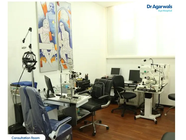
Clinical testing and workup
Black fungus diagnosis test include:
- Endoscopic examination of the nose
This is a black fungus diagnosis test includes a thin flexible tube with a tiny camera and light, called an endoscope is inserted into the nose. This allows the doctor to look at the nose and sinus passages.
- A biopsy of a swab taken from the nose
A swab is inserted into the nostril of the patient and rotated in place to obtain a sample of the tissue. This is then sent for examination under a microscope by a trained microbiologist. This examination can show the presence of the mold.
- CT / MRI scan
A CT or MRI scan may also be used to indicate certain changes that can indicate mucormycosis infection. This along with the clinical findings can help clinch the diagnosis.
Time is of utmost importance in the treatment of Mucormycosis and the investigative processes take no more than one day to produce reports.
- Black Fungus Treatment
The process of black fungal disease treatment is teamwork involving an ENT (Ear, nose, throat) specialist, ophthalmologist, neurologist and radiologist. If black fungal disease is suspected, the patient should receive medical attention at the earliest. Mucormycosis treatment at home should not be attempted without medical advice. Treatment for black fungus post diagnosis should happen at a medical center with advanced facilities.
For black fungal infection treatment, the ENT surgeon has to aggressively debride the necrotic or dead tissue from the nose and the sinus. In case, the eye is involved, then the fungal material from around the eye also has to be removed.
In other cases, where advanced black fungus treatment is required, the entire orbit or the space around the eye is also involved, the eye has to be removed in a process called orbital exenteration.
Be it the eye or upper jaw, these can be replaced with appropriate artificial substitutes or prostheses. While prosthetic replacement of the missing facial structures can commence once the patient stabilizes after surgery, it is important to reassure patients about the availability of such interventions instead of leaving them to panic with the sudden unforeseen loss, augmenting a post-Covid stress disorder which is already a reality.
Along with the surgery, treatment for black fungus will also include the administration of antifungal medication. The most commonly used medication is Amphotericin B. Initially, this medicine is infused intravenously and if the patient shows improvement, they can be shifted to oral antifungal medication.
Doctors will also treat the underlying risk factors that are associated with mucormycosis infection.
Black fungus treatment in advanced cases can lead to loss of the upper jaw and sometimes even the eye. Patients would need to come to terms with the loss of function due to a missing jaw — difficulty with chewing, swallowing, facial aesthetics, and loss of self-esteem.
Be it the eye or upper jaw, these can be replaced with appropriate artificial substitutes or prostheses. While prosthetic replacement of the missing facial structures can commence once the patient stabilizes after surgery, it is important to reassure patients about the availability of such interventions instead of leaving them to panic with the sudden unforeseen loss, augmenting a post-Covid stress disorder which is already a reality.
Mucormycosis (Black Fungus)
What is Black Fungus (Mucormycosis)? Black fungus, scientifically known as mucormycosis, is a rare but...
Learn more about Mucormycosis (Black Fungus)
THE SCIENCE BEHIND THE TECHNOLOGY
The success of Contoura Vision lies in its precision. Unlike conventional LASIK procedures that use standard measurements, Contoura Vision employs a sophisticated diagnostic technique known as topography-guided mapping. This involves creating a 3D map of the cornea’s imperfections, capturing even the most minute irregularities. With this highly detailed map, the laser can target specific imperfections with unparalleled accuracy.
By customizing the laser treatment to the unique topography of each patient’s cornea, Contoura Vision not only corrects refractive errors but also addresses higher-order aberrations that can significantly impact vision quality. This level of precision results in sharper, clearer vision, often surpassing what glasses and contact lenses can offer.
BENEFITS OF THE PROCEDURE
Contoura Vision offers a plethora of benefits that have made it a preferred choice for many seeking vision correction:
1. Sharper Vision:
Contoura Vision often provides patients with a vision that surpasses what they could achieve with glasses or contact lenses.
2. Customization:
Each procedure is tailored to the individual, addressing their unique corneal irregularities for optimal results.
3. Quick and Painless:
The procedure is quick, usually lasting only a few minutes, and patients typically experience minimal discomfort.
1. Fast Recovery:
Most patients notice improved vision within a day or two, and many can return to their normal activities shortly after the surgery.
2. Reduced Dependence on Glasses:
Many patients find that they no longer need glasses or contact lenses for daily activities after Contoura Vision.
3. Long-Lasting Results:
Contoura Vision results are generally stable over the long term,offering lasting benefits.
HIGHLIGHTS OF CONTOURA VISION PROCEDURE
- No pain
- No cut
- No stitches
- No hospitalization
- Quick recovery
CONTOURA VISION VS. TRADITIONAL LASIK
1. Precision:
Contoura Vision’s topography-guided mapping system provides a level of precision that traditional LASIK cannot match.
2. Visual Quality:
Contoura Vision often results in better visual quality, particularly in lowlight conditions and for those with higher-order aberrations.
3. Customization:
Traditional LASIK uses standardized treatments, while Contoura Vision tailors the procedure to each patient’s unique corneal topography.
4. Reduced Side Effects:
Contoura Vision has been associated with fewer side effects such as glare and halos compared to traditional LASIK.
CONTOURA VISION PROCEDURE – PROCESS
Contoura Eye surgery is increasingly becoming a popular vision correction treatment option for people all around the country. It is a quick, painless same-day surgery, helping you gain excellent vision in 15 to 20 minutes.
Pre-Operative Eye Exam
A comprehensive preliminary assessment is performed, including a clinical examination and corneal topography (Pentacam), to determine the patient’s eligibility and safety for the Contoura LASIK surgery. If any retinal defects (such as thinning/hole/tear) are discovered during the evaluation, they are treated with a barrage laser first, and then Contoura is performed after 1 to 4 weeks.
During the Procedure
On the day of surgery, the topolyser collects the patient’s topographic imagery, which is then sent to the treatment planning station to develop the personalized treatment profile. The laser will adjust your vision by accurately reshaping your cornea based on up to 22,000 elevation points, guided by your individualized topography profile.
Post Procedure
After 30 minutes, the patient is free to exit the hospital. Eye drops are used to prevent infection and aid in the healing process. The patient is evaluated the day following the procedure and one week and one month after that.
WHY CHOOSE CONTOURA
- US FDA-Approved
- Excellent night vision
- Quick results
- Walk-in and Walk-out spectacles free
If you’re tired of the limitations of glasses and contact lenses and dream of experiencing the world with clarity and freedom, Contoura Vision could be the solution you’ve been waiting for—a future where perfect vision is no longer a distant dream but a reality
Neuro Ophthalmology – When should you visit your eye doctor?
Neuro Ophthalmic issues are quite a concern for doctors; for if not treated on time, it could result in Optic Nerve atrophy (death of the optic nerve).
Some of the most common signs of Optic Nerve Dysfunction include:
- Reduced visual activity all of a sudden
- Double vision and headaches
- A less reactive pupil (pupil is the central part of eyeball that allows light to pass through)
- Impairment of colour vision (especially inability to identify red & green colours)
- Difficulty in seeing light (Photophobia)
- Visual Field Defects (visibility coverage)
Neuro Ophthalmic Conditions – And here come the ‘scary’ words
It’s quite natural that you’ll hear a lot of medical jargons in your doctor’s office. However, your doctor will try and explain the details, so you understand the illness/condition well and proceed with the available treatment options. Here are a few common conditions pertaining to neuro ophthalmology:
Optic Neuritis:
This is a condition that involves inflammation of the optic nerve. An inflammation could occur due to various reasons – starting from an infection to an autoimmune disorder.
Papilloedema:
In this case, the optic disc (the circular area where the optic nerve connects to the retina, at the back of the eye) swells up due to an excessive pressure from inside the skull may be due to a tumor for instance.
Nutritional Optic Neuropathy:
Here the damage to the optic nerve is caused by certain toxic substances found in tobacco & alcohol. This could also occur due to lack of nutrients and deficiency of vitamin B-complex and folic acid.
Diabetic Neuropathy:
In this, the optic nerve is damaged due to the excessive blood sugar or diabetes. As the disease progresses, the blood supply to the retina gets cut-off, leading to vision loss.
Although each of them attacks the optic nerve in a different way, the final outcome is eventually the death of the optic nerve, if left untreated.
Neuro Ophthalmology Treatments – Pills or prayers?
Your eye specialist will ideally be able to pick up an optic nerve issue during an eye examination. A brain CT scan or an MRI is then typically done to assess whether there is associated damage in the brain as well. While some conditions can be treated with oral medications and injectables, others might require a surgery. Some of the common treatment options would be:
Optic Neuritis:
Antibiotics and corticosteroids are used to flush out the infection or suppress the immune disorders from causing further damage.
Papilloedema:
Medications are used to reduce the pressure inside the skull. Might require a brain surgery if the increased pressure is due to a tumorous growth.
Nutritional Optic Neuropathy:
Lifestyle changes are recommended and vitamin injectables are prescribed.
Diabetic Retinopathy:
Since the underlying cause is diabetes, it is essential to treat the diabetes before it further damages the nerve and other blood vessels.
If not detected on time, there’s a good chance for complete loss of vision.
Written by: Dr. Preetha Rajasekaran – Consultant Ophthalmologist, Porur
One day, imagine experiencing a clear eyesight that you haven’t had in years. These days, a lot of people with eye diseases like keratoconus or corneal ectasia can actually achieve this because of breakthroughs in eye surgery. A notable example of this is the CAIRS eye surgery. If you or a loved one is thinking about having this operation done, we’ll walk you through everything you need to know, making sure you understand every step and feel confident.

How Does CAIRS Treatment Procedure Work?
With keratoconus, a progressive eye disease, vision becomes distorted as the cornea thins and attains cone-shaped. In order to stabilise and restructure the cornea, corneal ring segments are implanted during the CAIRS operation. The below four points will guide you about thorough rundown of the CAIRS treatment process:
1. Indications
CAIRS is often recommended for patients with:
- Progressive Keratoconus.
- Other corneal ectasias that do not respond well to conservative therapies, such as rigid contact lenses.
- Patients who are not good candidates for corneal collagen cross-linking or other surgical procedures.
2. Preoperative Assessment
Before the surgical treatment, a comprehensive eye examination is performed as follows,
- Corneal Topography is used to map the corneal shape and assess the extent of the ectasia.
- Pachymetry is used to determine corneal thickness.
- Ocular History and Visual Acuity Testing are used to determine the influence on vision and set a baseline.
- The purpose of the contraindications evaluation is to ensure that no conditions, such as active infection or extensive corneal scarring, preclude surgical intervention.
3. CAIRS Procedure
Anaesthesia
- The surgical procedure is usually done under local anaesthesia with topical anesthetic drops.
Creation of Stromal Tunnel
- A femtosecond laser or a mechanical microkeratome is utilised to create a precise tunnel through the corneal stroma. This tunnel is where the corneal segments will be inserted.
- The depth and length of the tunnel are carefully estimated using preoperative measurements.
Preparation of Allogenic Segments
- CAIRS corneal segments are created from the donor corneal tissue. These segments are formed into little rings or arcs that offer structural support for the cornea.
- To assure its suitability for implantation, allogenic tissue is treated and sterilised.
Insertion of Segments
- The allogenic corneal ring segments are carefully placed in the stromal tunnel.
- The positioning is critical for producing the desired effect on corneal shape and stability. The severity and asymmetry of the keratoconus determine whether one or two segments are inserted.
Final Adjustments and Healing
- Following insertion, the segments are adjusted to ensure optimum alignment and positioning.
- Antibiotic and anti-inflammatory drops are given to help prevent infection and inflammation.
4. Postoperative Care
- Patients are carefully monitored following surgery, with regular follow-up sessions.
- They are given a regimen of antibiotic and anti-inflammatory eye drops.
- Visual acuity and corneal topography are examined on a regular basis to ensure the procedure’s success and detect any problems at the earliest.
Benefits of CAIRS for Keratoconus
The CAIRS technique has many benefits for individuals with keratoconus, making it a viable choice for managing this degenerative eye disease. Here are the main benefits of CAIRS for keratoconus:
1. Stabilisation of Corneal Shape
- CAIRS gives structural support to the cornea, slowing the course of keratoconus by preventing additional thinning and bulging.
- The use of allogenic segments can result in long-term stabilisation of the corneal shape, eliminating the need for future invasive treatments.
2. Improvement in Vision
- By reshaping and stabilising the cornea, CAIRS can considerably reduce irregular astigmatism, a prominent source of visual distortion in keratoconus patients.
- Many patients report better visual acuity as the corneal shape becomes more regular, resulting in clearer and sharper eyesight.
3. Minimally Invasive Procedure
- CAIRS is less invasive than standard corneal transplantation (penetrating or deep anterior lamellar keratoplasty), which requires more intensive surgery and a longer recovery time.
- It often has a quicker recovery period than more invasive surgical treatments, allowing patients to resume their regular activities sooner.
4. Compatibility with Other Treatments
- CAIRS can be used in conjunction with corneal collagen cross-linking (CXL), which strengthens the corneal collagen fibres. The combination can improve stability and vision.
- It can be adjusted to specific patient demands by adjusting the number and location of the ring segments based on keratoconus severity and asymmetry.
5. Use of Donor Tissue
- The use of allogenic (donor) corneal tissue segments assures greater biocompatibility and lowers the likelihood of adverse responses when compared to synthetic implants.
- Donor tissue blends seamlessly with the patient’s cornea, facilitating natural healing and lowering the risk of rejection or extrusion.
6. Potential for Delay or Avoidance of Transplantation
- By stabilising the cornea early in the disease process, CAIRS can postpone or even eliminate the need for corneal transplantation, a more complicated and hazardous treatment.
- Delaying or postponing transplantation can also save money in the long run, lowering the patient’s overall healthcare burden.
7. Customizability
The technique can be customised to the patient’s individual corneal shape and degree of ectasia. Surgeons can modify the number, size, and positioning of the segments to produce the best results.
Who Is Required to Do This CAIRS Procedure?
The CAIRS procedure should be conducted by a highly skilled eye surgeon with a medical degree and ophthalmology residency. Ideally, the surgeon should have extra fellowship training in cornea and refractive surgery, which allows for specialised competence in treating corneal illnesses and executing advanced corneal procedures. They must be board-certified in ophthalmology and have extensive experience diagnosing and managing keratoconus, as well as knowledge of corneal surgical methods, particularly those using intrastromal implants.
Experience with modern equipment such as femtosecond lasers or mechanical microkeratomes is also required. To guarantee comprehensive patient care, the surgeon should participate in ongoing education to stay up to date on the newest breakthroughs, join relevant professional organisations, and collaborate with a multidisciplinary team of experts. To achieve the best results, effective communication skills are required while meeting with patients, explaining the operation, and giving detailed postoperative care.
Will I See Better After Having CAIRS Surgery?
Many individuals see significant improvements in their vision after CAIRS surgery, while the level of improvement varies depending on the severity of the keratoconus, prior eyesight, and corneal features. It can decrease irregular astigmatism and increase visual acuity, resulting in crisper and sharper vision. Patients frequently report better eyesight, with fewer distortions and glare. The surgeon’s precision in segment placement, adherence to postoperative care recommendations, and corneal health all contribute to the surgery’s outcome. While CAIRS primarily tries to stabilise the cornea and slow disease progression, many patients still require corrective lenses, albeit less strong ones. It is critical to set reasonable expectations and explore possible outcomes with the surgeon.
Is CAIRS the Only Option Left to Improve My Sight, or Are There Other Treatments?
CAIRS is one of various methods for improving eyesight in people with keratoconus and other corneal ectatic conditions. Other eye treatments include glasses and contact lenses, which can correct vision in the early stages; rigid gas permeable (RGP) and scleral lenses, which provide a more consistent refractive surface for moderate to advanced keratoconus; and corneal collagen cross-linking (CXL), which strengthens the corneal collagen fibres and slows disease progression. Furthermore, Intacs (intrastromal corneal ring segments) are synthetic implants used to reshape and stabilise the cornea, similar to CAIRS but with plastic segments instead of donated tissue. The severity of the condition, corneal characteristics, and unique patient demands all influence treatment decisions, which frequently necessitate a consultation with a trained ophthalmologist to establish the best strategy.
How Much Does CAIRS Surgery Cost?
In India, the cost of CAIRS varies based on the patient’s eye features and the type of corneal problem being treated. The severity of keratoconus, as well as the particular corneal shape and thickness, can all have an impact on the procedure’s difficulty and cost. Furthermore, the geographic location, surgeon expertise, and kind of medical facility all have an important impact in deciding the final keratoconus surgery cost. A full consultation with an experienced ophthalmologist is required to provide an accurate cost estimate based on your specific needs and corneal health.
Who Developed the CAIRS Procedure?
Dr. Soosan Jacob, a distinguished ophthalmologist and pioneer in corneal and refractive surgery at Dr Agarwals Eye Hospital, created the CAIRS procedure. Dr. Soosan Jacob is well-known for her unique contributions to ophthalmology, which have helped advance numerous surgical approaches for treating difficult corneal problems. Her CAIRS method, which uses allogenic tissue to stabilise and restructure the cornea, is a revolutionary strategy for treating keratoconus and other corneal ectatic problems.
Verified by: Dr. T. Senthil Kumar MBBS MS (Ophthal) (Gold Medallist) FICO
Reference:
- Jacob S, Agarwal A, Awwad ST, Mazzotta C, Parashar P, Jambulingam S. Customised corneal allogenic intrastromal ring segments (CAIRS) for keratoconus with decentered asymmetric cone. Indian Journal of Ophthalmology/Indian Journal of Ophthalmology. https://pubmed.ncbi.nlm.nih.gov/37991313/
How is the penetrating keratoplasty surgery performed?
The penetrating keratoplasty surgery is usually performed under local anaesthesia. Through a small corneal incision (opening), the endothelium is removed from the patient’s eye and a disc of donor endothelium is inserted in the patient’s eye which is placed in position with the help of an air bubble.
A few stitches may be taken which will be removed 3-4 weeks after the surgery. Once keratoplasty surgery is over, the patient needs to lie down flat for a few hours for proper attachment of the graft. The air bubble usually gets absorbed in 48 hours but may take longer.
What are the indications of Penetrating Keratoplasty (PDEK)?
- Fuch’s endothelial dystrophy
- Pseudophakic bullous keratopathy
- Aphakic bullous keratopathy
- ICE syndrome
Endothelial dysfunction secondary to glaucoma
What are the advantages over full thickness penetrating Keratoplasty?
- Few sutures required as compared to penetrating keratoplasty.
- Suture induced astigmatism is avoided
- Suture related complications are avoided
- Greater stability
- Faster visual rehabilitation
- The graft can be obtained from any age group of the donated eyes
- Chances of rejection are less
What are the complications of Penetrating Keratoplasty (PDEK)?
- Graft detachment/ dislocation
- Recurrent epithelial erosions
- Cataract formation
- Glaucoma
- Graft rejection
- Graft failure
What is corneal graft rejection?
The donor eye is genetically different from the patient’s body, due to which the patient’s body tries to fight against it. This is called corneal graft rejection.
What are the symptoms of corneal graft rejection?
The symptoms are: Redness, Sensitivity to light, Vision drop, Pain (RSVP). Along with sticky discharge and foreign body sensation.
Report to your Ophthalmologist as soon as possible if any of the above symptoms show up post-surgery.
How do I prevent graft rejection?
- In order to prevent rejection, a list of anti-rejection medications will be prescribed by your doctor, which should be used religiously.
- You should have an adequate supply of eyedrops at home so that a single dose is not missed.
- Do not stop any medication without consulting your Ophthalmologist.
- In case any of the above symptoms of rejection occur, meet your Ophthalmologist immediately. It can often be reversed if anti-rejection medications are started promptly. It should also be noted that rejection can occur at anytime in the years to come.
- Review regularly to check for vision, intraocular pressure, graft condition and retinal assessment.
What are the risk factors for rejection?
- Vascularisation over the cornea
- Pre-existing inflammation
- Corneal transplant done more than 2 times
- Pre-existing glaucoma
What is graft failure?
When the corneal graft rejection has not been promptly treated or does not respond to anti- rejection medication, graft failure has occurred. The only way of managing graft failure is by replacing the graft. In addition, there are three types of graft rejection: acute, hyperacute, and chronic rejection.
Written by:Dr. Preethi Naveen – Training Committee Chair – Dr. Agarwals Clinical Board
Principle
A pinhole or a small aperture is created, thereby allowing passage of rays of light from the central aperture and blocking the rays emanating from the peripheral irregular cornea, so that the impact of higher order aberrations caused by irregular corneal astigmatism can be minimized. Another mechanism is the Stiles-Crawford effect of the first kind, according to which, an equal intensity of light entering near the centre of the pupil produces a
greater photoreceptor response compared with the light entering the eye near the edge of the pupil. Therefore, when the pupil narrows, more focused light enters the eye through the narrow aperture, producing a greater photoreceptor response.
Procedure
- Under peribulbar anaesthesia ,4 mL lidocaine hydrochloride (Xylocaine 2.0%) and 2 mL bupivacaine hydrochloride 0.5% (Sensorcaine)
- 2 paracenteses are created and a 10-0 polypropylene suture attached to the long arm of the needle is introduced into the anterior chamber.
- The anterior chamber can be maintained with an ophthalmic viscosurgical device or with fluid infusion with the help of an anterior chamber
- maintainer or a trocar anterior chamber maintainer.
- An end-opening forceps is introduced through the paracentesis, and the proximal iris leaflet is held. The suture needle is passed through the
- proximal iris tissue.
- A 26-gauge needle is introduced from the paracentesis from the opposite quadrant and passed through the distal iris leaflet after being held with end-opening forceps. Next, the tip of the 10-0 needle is then passed through the barrel of the 26-gauge needle, which is then pulled out of the paracentesis. The 10-0 needle exits the anterior chamber along with the 26-gauge needle.
- A Sinskey hook is passed through the paracentesis, and a loop of suture is withdrawn from the eye. The suture end is passed through the loop 4 times. Both the suture ends are pulled and the loop slides inside the eye, approximating the iris tissue edges. The suture ends are then cut with micro scissors and the procedure is repeated in the other quadrant to achieve a pupil of desired configuration and to decrease the pupil to pinhole size.
Indications
- Functional or Optical:
Symptomatic iris defects (Congenital, Acquired, Iatrogenic, Traumatic)
- Oppositional angle closure or PAS:
To break PAS and angle apposition angle closure glaucoma whether primary, post trauma, plateau iris
syndrome, Urrets-Zavalia syndrome or long-standing silicone oil in the anterior chamber.
- Cosmesis:
PPP can be done for cosmetic indication, especially in large colobomas.
- Penetrating Keratoplasty:
In cases of floppy iris that is expected to adhere to the peripheral edge of graft causing peripheral anterior synechiae,
pupilloplasty is performed to tighten the iris preventing it from causing synechial adhesions that would increase the risk of angle closure and graft failure.
Advantages
- Faster and easier to perform compared to other pupilloplasty techniques – (Modified Siepser’s and McCanell method which requires more than two passes to be made from the anterior chamber, as well as additional manipulation of the iris tissue).
- Reduced postoperative inflammation and faster visual recovery
- Effective in Urrets Zavalia syndrome who present with raised IOP and persistent pupil dilation.
- Prevents secondary angle closure, breaking the formation of peripheral anterior synechia and inhibits mechanical blockage.
- Useful in treating patients with higher order corneal aberrations, improves visual quality and extended depth of focus.
- Effective in selected cases of secondary angle closure, along with silicon oil induced glaucoma.
- Reconstructing the pupil this way prevents patients from glare, photophobia and untoward images formed by reflection of light
Disadvantages
- Limited dilation- to examine the posterior segment – (In cases of retinal detachment, it is possible to YAG the iris and undo procedure if needed).
- Chances of touching crystalline lens during procedure and risk of cataract formation – So preferably done in pseudophakic eyes.
Written by: Dr. Soundari S – Regional Head – Clinical Services, Chennai
Paediatric Ophthalmology – Problems affecting our little people
Research shows that 1 out of 6 kids has vision related issues. Some of the most common issues affecting the little ones include:
- Squint
- Drooping of upper eyelids
- Lazy eye
- Refractive-errors
Eye diseases in newborns include:
It is essential to address and rectify the issues in newborns as soon as possible. If not treated within the first six months of the child’s birth, there’s a good possibility that the child gets visually impaired for the rest of his/her life. The reason being, the optic nerve connecting the eyes to the brain is still developing and if any prevalent disease is not treated on time, there could be a permanent disconnect between the eyes and brain, eventually leading to total blindness.
Paediatric Ophthalmology – Let’s nip it in the bud!
Routine comprehensive eye checks should be an essential part of your child’s healthcare regime. While problems like squint or drooping of eyelids can be easily noticed, finding issues related to lazy eye & refractive errors could be quite a challenge to the parents. Especially because most kids do not report the problem to their parents for often they lack the ability to understand that there’s been a change in their visual skills. It, therefore, becomes the primary responsibility of parents to notice any change in their kids’ behavioural pattern like watching TV from a close distance or excessively straining to read from a book or performing badly at school all of a sudden.
If any of these ring a bell, then it’s time to meet a paediatric ophthalmologist and clarify on your kid’s eye health.
Paediatric Ophthalmology – Saving the vision of our tomorrow
Paediatric Ophthalmology is taken quite seriously at Dr. Agarwal’s Eye Hospitals with expert consultants & surgeons working round-the-clock to make sure, the vision of our future generation is well protected. Children with squint and lazy eye issues are initially treated by prescribing glasses and suggesting eye exercises. In fact, Dr. Agarwal’s was one of the first hospitals to introduce the concept of eye yoga as a treatment mechanism. Parents of children born from a wedlock between relatives or with both of them wearing glasses due to refractive errors are advised to bring their kids for an evaluation, right from 3-4 years of age.
Black Fungus (Mucormycosis) Symptoms in the Eye
Early detection of black fungus in eyes is essential to prevent vision loss or severe complications. Symptoms may include:
1. Swelling and Redness Around the Eye
Mucormycosis can cause inflammation around the affected eye, leading to visible swelling and redness.
2. Blurred or Double Vision
Patients may experience black fungus symptoms such as blurry or double vision due to the infection spreading into the eye socket.
3. Eye Pain and Sensitivity to Light
Eye discomfort, along with increased sensitivity to bright light, is another key indicator.
4. Dark Patches Around the Nose and Eyes
A distinctive sign of black fungus infection is the presence of darkened skin patches near the eyes and nasal area, caused by dead tissue.
5. Partial or Complete Vision Loss
In severe cases, the fungal infection may damage the optic nerve, leading to irreversible vision loss if left untreated.
Causes of Mucormycosis or Black Fungus
Understanding the causes of mucormycosis can help in early prevention and treatment. The primary causes include:
1. Weakened Immune System
Individuals with compromised immunity, such as those undergoing chemotherapy or organ transplants, are at a higher risk of developing mucormycosis disease.
2. Uncontrolled Diabetes
Diabetes, particularly uncontrolled blood sugar levels, creates an environment where fungi thrive, increasing the chances of infection.
3. Prolonged Use of Steroids
The excessive use of steroids, often prescribed for severe respiratory illnesses, can lead to black fungus infection due to immune suppression.
4. Exposure to Mold Spores
Molds responsible for mucormycosis are present in soil, decaying plants, and dust. Inhaling these spores can result in fungal infections in vulnerable individuals.
5. Contaminated Oxygen Support Equipment
During the COVID-19 pandemic, cases of black fungus were linked to unclean oxygen cylinders, humidifiers, and medical equipment.
Symptoms & Causes
Black Fungus Symptoms & Causes
Mucormycosis is caused by exposure to mucor mold which is commonly found in soil, plants, manure,...
Risk Factors of Black Fungus Infection
Certain individuals are more prone to developing mucormycosis. Risk factors for black fungus infection include:
- Diabetes and high blood sugar levels
- Extended steroid use (especially during COVID-19 treatment)
- Cancer or undergoing chemotherapy
- Organ transplants or weakened immunity
- Poor hygiene and environmental exposure
How to Prevent Black Fungus (Mucormycosis)?
While mucormycosis is a severe condition, preventive measures can reduce the risk:
- Maintain proper hygiene: Keep nasal passages clean and avoid dusty environments.
- Monitor blood sugar levels: Diabetic patients should manage their glucose levels effectively.
- Use steroids cautiously: Only take steroids as prescribed by healthcare professionals.
- Avoid prolonged exposure to moldy environments: Stay away from damp or decaying surroundings.
- Strengthen immunity: A balanced diet, regular exercise, and adequate sleep support immune health.
Types of Mucormycosis or Black Fungus
There are different types of mucormycosis, classified based on the affected area of the body:
1. Rhinocerebral Mucormycosis (Sinus and Brain)
This form affects the sinuses and brain, often spreading from the nasal cavity. Symptoms include severe facial pain, headache, nasal congestion, and blackened nasal tissue.
2. Pulmonary Mucormycosis (Lungs)
Common in individuals with lung disease or those undergoing chemotherapy, this type presents with fever, chest pain, and difficulty breathing.
3. Cutaneous Mucormycosis (Skin and Wound Infections)
Affects the skin following surgery, burns, or injuries, causing redness, swelling, and painful ulcerations.
4. Gastrointestinal Mucormycosis
This type occurs in the digestive tract and is often found in premature infants or individuals with malnutrition.
5. Disseminated Mucormycosis
The most severe form, where the infection spreads throughout the bloodstream, affecting multiple organs, including the brain.
When to See a Doctor for Black Fungus (Mucormycosis) Disease?
Seek medical attention if you experience:
- Persistent facial pain or swelling
- Black patches around the nose or eyes
- Difficulty breathing or chest pain
- Sudden vision changes or eye swelling
- Unexplained headaches and fever
Early diagnosis is crucial in managing mucormycosis treatment effectively and preventing life-threatening complications. Mucormycosis is a severe fungal infection that requires immediate attention. Early detection, proper hygiene, and timely medical intervention can help prevent its devastating effects. If you notice any black fungus symptoms, consult a healthcare provider immediately.
Treatment of Black Fungus (Mucormycosis)
The treatment of black fungus mucormycosis involves antifungal medications and, in some cases, surgical procedures to remove infected tissue. Common treatment options include:
- Amphotericin B: A strong antifungal drug administered intravenously.
- Posaconazole or Isavuconazole: Oral antifungal medications used for long-term treatment.
- Surgical Removal of Affected Tissue: In severe cases, surgical debridement is necessary to prevent the spread of infection.
Black Fungus Treatment & Diagnosis
Black fungus diagnosis is challenging because the symptoms are common to several other conditions It s diagnosis hence involves a...
Why Does Eyes Need Moisture?
Adequate quality and quantity of tears in the eyes is essential for smooth feeling and functioning of the eyes.
The dry eye syndrome is the change in quality or quantity of the three layers of the tear film – Oily (exterior), water/aqueous layer (middle) and protein (inner).
What are the Common Causes of Dry Eye?
Commonest cause of dry eyes are the air conditioners. The artificial air and temperature change caused by the air conditioners can cause changes and effect the largest organ of the body- the skin to the immune system, to the most delicate organ the eyes. The extreme loss of humidity and resultant dryness in the air around us especially in low temperature settings in AC, causes more evaporation from the watery layer of tear film , causing evaporative dry eyes and subsequently, long term exposure to such AC can also alter the lipid production from glands in the eyelids causing both change in quality and quantity of tear film and hence dry eyes.
The tears have antimicrobial functions and in dry eyes, when there is no adequate lubrication, the eyes are also more vulnerable to inflammation and infections which could lead to decrease in vision.
The other important causes of dry eye disease are:
- Prolonged staring/usage of computer/mobile phones (Computer Vision Syndrome).
- The natural aging process, especially menopause problems and hence women are more affected by dry eyes .
- Certain medical conditions including diabetes, thyroid disorders and Vitamin A deficiency
- Side effects of certain drugs like antihistamines can also be a cause of dry eye.
- Laser eye surgery, though symptoms of dry eyes related to this procedure are usually temporary.
- Tear gland damage from inflammation or radiation
- Diseases that affect your ability to make tears, like Sjogren’s syndrome, rheumatoid arthritis, and collagen vascular diseases
- Problems that don’t allow your eyelids to close the way they should.
- Air pollution -people in metros like New Delhi, Mumbai, Kolkata and Chennai are few times more likely to be diagnosed with dry eye syndrome than the other towns with relatively little air pollution.
Major symptoms of dry eye disease:
Symptoms of dry eyes and dry eye syndrome can be Burning ,dryness ,grittiness, Itchiness, Aching sensations, Heaviness , watering from the eyes and blurred vision. The reading speed can slow down for dry eyes and the rate decreases as the severity increases.
Tips & Treatment to Prevent Symptoms of Dry Eye Syndrome:
- Try to minimize and decrease the number of hours of using air-conditioned rooms, setting of AC temperature around 23 degree C and above is recommended.
- Avoid sitting with your face facing air conditioners, thereby preventing eyes from being directly exposed to air from air-conditioners.
- Place a small open bowl of fresh water in the corner of the room you sit with the air conditioner to maintain the humidity of the room, thereby preventing dry skin and dry eyes. So far, this has proven to be one of the best dry eye remedies.
- Another dry eyes home remedy is to drink enough liquids can also help prevent dry eye syndrome.
- Frequent conscious blinking when using computer or mobile phones which will help in the appropriate distribution of tear film.
- Have a traditional 7 – 8 hours of sleep, which will give adequate rest to the eyes.
- Consider wearing sunglasses or protective eye wear.
- Reach out to your eye doctor to diagnose the cause of your dry eyes, and follow recommendations and prescriptions from the Eye doctor on eye medications like lubricants, and /or antibiotics and/or anti-inflammatory eye drops and other in-office procedures which could help your body create and secrete more tears and better quality of tears and to decrease eye irritation and inflammation.
If precautionary methods are not followed to prevent symptoms of dry eye syndrome, people will also develop eye infections. In the case of untreated, dry eyes, as the severity and duration of dry eyes increase, patients might develop damage to the corneal surface (abrasion), corneal ulcer and serious vision problems.
Prevention is better than cure. Get your eyes checked for dry eyes syndrome and get them treated appropriately.
Types and benefits of Retina Laser
According to the type of retinal disorder, laser therapy is provided in different ways.
Proliferative Diabetic Retinopathy (PDR)
- Proliferative diabetic retinopathy is a form of advanced or end-stage diabetic retinopathy. Due to the long duration of diabetes and uncontrolled blood sugar levels, the retinal blood vessels undergo changes which happen in stages, ultimately leading to PDR. PDR is a vision-threatening disorder. When timely treatment is not provided, it can cause complications like bleeding within the eyes from the abnormal vessels and/or can retinal detachment.
- Retinal laser therapy is helpful in PDR as it decreases the risk of such complications. The doctor performs pan-retinal photocoagulation (PRP) to treat PDR.
- The retina is a 360-degree structure that is responsible for vision. The central retina is called as macula and is the chief zone responsible for fine vision. During proliferative diabetic retinopathy, the doctor applies laser therapy to the poorly vascular retinal areas sparing the macula. Proliferative diabetic retinopathy therapy is provided in three to four sessions since the almost 360-degree retina is slowly covered with laser spots. The formation of abnormal blood vessels and undue complications are prevented by this procedure.
Diabetic Macular Edema (DME)
DME is abnormal fluid collection leading to swelling at the level of the macula, causing vision loss. Retinal laser photocoagulation is beneficial in some cases of DME. Here, minimal laser spots are given targeting the leaky macular blood vessels to reduce the swelling.
Retinal Vein Occlusion (RVO)
In RVO, the entire retinal vessel or a part of the retinal vessel gets blocked due to various reasons leading to abnormal blood flow to the part of the retina supplied by the vessel. Here, Retinal laser therapy is useful, similar to PRP in PDR, as explained before.
Retinal Tears, Holes and Lattice Degeneration
Retinal tears, holes and lattice degenerations (areas of retinal thinning) occur in almost 10% of the normal population and are more common among myopes. If not treated, there is always a risk of developing retinal detachment through the breaks.
The doctor, in such cases, can delimit the retinal breaks with two to three rows of laser spots around the breaks, thus causing dense adhesion in the surrounding retina and thereby decreasing the risk of retinal detachment. It is mandatory to screen and laser such lesions prior to LASIK and cataract surgeries.
Central Serous Chorioretinopathy (CSC) and Choroidal Neovascularization
Both the conditions lead to areas of leak at the macular level, causing fluid collection and vision loss. Based on the specialist’s decision, in some cases, retinal laser therapy targeting the leaky areas is beneficial.
Patient preparation
The laser procedure is performed only after providing topical anaesthesia. Eye drops would be used prior to the procedure to minimize pain. The procedure is relatively painless. The patient might feel a mild pricking sensation during the therapy. The entire procedure might take place for five to twenty minutes, depending on the patient’s disease.
After the procedure
The patient might feel mild glare and visual discomfort for a day or two. He or She will be advised to use antibiotic and lubricant eye drops for 3 to 5 days, depending on the type and duration of the procedure. Extensive PRP in diabetic retinopathy can lead to a decrease in contrast sensitivity and colour vision.
Types and method
There are two methods by which laser therapy can be performed: Contact and Non-Contact methods. In the contact procedure, a lens with a lubricating gel will be placed over the patient’s eyes, and laser therapy would be delivered in sitting position.
In the non-contact method, the patient is made to lie down, and laser therapy is delivered. Sometimes the doctor might apply minimal pressure around the patient’s eyes with a handheld instrument.
Conclusion
Retinal laser photocoagulation is a relatively safe, fast and a painless procedure.
Written by: Dr. Dheepak Sundar – Consultant Ophthalmologist, Velachery
What are the retinal diseases which can be treated with cryotherapy?
Retinal tears to prevent retinal detachment, to seal leaking blood vessels, to slow or stop the growth of abnormal blood vessels caused by diabetic retinopathy
How does cryopexy help in treating retinal disorders?
This treatment creates a scar around retinal tears, around the abnormal blood vessels in order to stop the progression of the abnormal growth.
What are the precautions to be taken before the procedure?
It is an outpatient procedure. There are no special preparations for this procedure. You should eat normally and take all your regular medicines before you come in for the procedure
How is cryotherapy performed?
Cryopexy is administered with local anaesthesia to prevent pain. During the process, your ophthalmologist will use an indirect ophthalmoscope to view the inside of your eye through the pupil while gently pushing on the outside of the eye with the small metal probe to find the exact place for treatment. Once the appropriate treatment location is found, your doctor will activate the probe to deliver the freezing gas, which rapidly freezes the targeted tissue. As the tissue heals, it forms a scar.
What are anti VEGF Agents
Anti Vascular endothelial Growth factor (anti VEGF) agents a group of medications which block the activity of VEGF and thus mitigate the abnormal effects of VEGF
How have anti VEGF treatment influenced the management of various eye conditions
Anti VEGF agents when administered under appropriate conditions act at molecular level countering the action of VEGF and thereby reducing the morbidity.
Many diseases which were considered untreatable earlier like age related macular degeneration are rendered treatable, enabling patients maintain quality vision and subsequent improvement in quality of life
Ocular manifestation of systemic diseases with diabetes hypertension are also now treated with anti VEGF agents, with quality vision being restored and maintained.
How do I choose the type of anti VEGF agent
- The doctor examining you will prescribe the appropriate agents as per the disease process and systemic illness. Active bleeding or fluid leak at the back of the eye called macula warrants urgent treatment. The doctor will perform appropriate scans to confirm, quantify and monitor the progress of the disease. Vision is measured and is one of the yardsticks for monitoring response to treatment
How is the anti-VEGF agent administered
- After clinical examination and relevant scans and making a diagnosis, the doctor will discuss the available options with the patient
- The anti-VEGF agent is administered into the eye by means of a fine needle under sterile conditions in an operation theater.
- The eyes are numbed with topical anaesthetic agent
- Cleaning of eyes and surrounding structures with an antiseptic solution is done
- Protective sheet called eye drape is applied around the eye
- Eyelids are opened with a clip called an eyelid speculum
- The doctor injects the drug through the white part of the eye through a fine needle
- After injection, gentle massage is done at the site of injection
- The eye clip is removed, and antibiotic drops are instilled in the eye
Antibiotic drops are prescribed for usage after injection in the eye.
What are the anti-VEGF agents available for treatment?
- Bevacizumab
- Ranibizumab
- Aflibercept
- Brolucizumab
Written by: Dr. Mohanraj – Consultant Ophthalmologist, Coimbatore